Artery branches
- The AICA and the PICA give rise to the choroidal arteries, which supply the tela choroidea and attached choroid plexus.
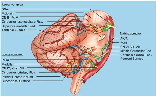
- Is supplied by three pairs of cerebellar arteries
Superior cerebellar artery SCAs (yellow)
- A branch of basilar artery
- Supplies the superior surface of the cerebellum.
- Arises at the midbrain level and encircles the brainstem near the pontomesencephalic junction.
- SCA courses below the oculomotor and trochlear nerves and above the trigeminal nerve.
- The SCA loops down closer to the trigeminal nerve in B than in A.
- Related to the
- Superior half of the 4th ventricular roof
- Cerebellomesencephalic fissure
- Course:
- Arise from Basilar → Pass around the midbrain above the trigeminal nerve and divide into rostral and caudal trunks → The branches loop deeply into the cerebellomesencephalic fissure → branches give off the precerebellar arteries, which pass along the superior cerebellar peduncles to the dentate nuclei.
Anterior inferior cerebellar artery AICAs (orange)
- A branch of basilar artery
- Supplies the anterior part of the inferior surface of the cerebellum.
- Arises at the pontine level and courses by the abducens, facial, and vestibulocochlear nerves.
- In A, both AICAs pass below the abducens nerves.
- In B, the left abducens nerve passes in front of the AICA and the right abducens nerve passes behind the AICA.
- Related
- Cerebellopontine fissures
- Lateral recesses
- Course:
- Arise from the basilar artery → pass near or between the CN7/8 + closely related to the cerebellopontine fissures, the flocculi, and the lateral recesses → AICAs divide into rostral and caudal trunks before reaching the CN7/8. →
- Rostral trunk passes between the nerves and along the middle cerebellar peduncle near the cerebellopontine fissure.
- Caudal trunk passes below the nerves and near the lateral recess to supply the lower part of the petrosal surface.
Posterior inferior cerebellar artery PICAs (red)
- A branch of vertebral artery
- Supplies the posterior part of the inferior surface of the cerebellum
- Arise from the vertebral artery at the medullary level
- Course in relation to the glossopharyngeal, vagus, accessory, and hypoglossal nerves.
- Related
- Caudal half of the roof
- Cerebellomedullary fissure.
- Course:
- Arise from the vertebral arteries → pass between the CN 9, 10, 11 → reach the cerebellomedullary fissure → Pass near the caudal pole of the tonsils, form the caudal loop → ascend through the cerebellomedullary fissure → intimately related to the caudal part of the ventricular roof → pass around the rostral pole of the tonsil form the cranial loop → pass through the telovelotonsillar cleft → They give off branches to the dentate nuclei near the superior pole of the tonsils.
- In the PICA course around the tonsils, they divide into medial and lateral trunks
The origin of the SCAs are quite symmetrical from side to side. There is slight asymmetry in the level of origin of the AICAs and marked asymmetry in the level of the origin of the PICAs, especially in A.
Relationships of the cerebellar arteries
Relationships with brainstem, and cerebellar-brainstem fissures.
- The SCA passes around the midbrain to enter the cerebellomesencephalic fissure, where it sends perforating branches into the posterior midbrain below a line between the superior and inferior colliculi, and down the superior peduncle to the dentate nucleus.
- The AICA loops around the flocculus and the facial and vestibulocochlear nerves.
- The left PICA passes between the rootlets of the nerves entering the jugular foramen and turns caudally around the lower pole of the left tonsil, which has been removed, and then ascends to form a cranial loop at the upper pole of the tonsil bordering the inferior half of the ventricular roof.
- The left half of the cerebellum has been removed. The SCA passes around the midbrain below the PCA in the lower part of the ambient and quadrigeminal cisterns, enters the cerebellomesencephalic fissure, and loops over the posterior lip of the fissure to supply the tentorial surface.
- The PICA arises from the vertebral artery, passes around the medulla, crosses the inferior cerebellar peduncle, and enters the cerebellomedullary fissure, where it passes along the inferior half of the ventricular roof, and exits the fissure to supply the suboccipital surface. The AICA passes laterally around the pons and above the flocculus.
- The right PICA loops around the caudal and rostral poles of the tonsil. The left PICA dips below the level of the foramen magnum.
- The peak of the 4th ventricle
- The superior cerebeller peduncle white matter tracts arise from dentate nucleus which is where dentate nucleus is located
- The SCA passes above the trigeminal nerve and enters the cerebellomesencephalic fissure, where it sends branches down the superior peduncle to the dentate nucleus.
- The flocculus and choroid plexus project laterally from the margin of the foramen of Luschka into the cerebellopontine angle, behind the glossopharyngeal and vagus nerves and above the PICA.
- The hypoglossal rootlets arises from the medulla in front of the glossopharyngeal and vagus nerves and cross the posterior surface of the vertebral artery. Some hypoglossal rootlets pass above and others below the PICA origin.
SCA relationships
- The right SCA arises from the basilar artery as a duplicate artery.
- The rostral duplicate trunk gives rise to vermian branches that supply the vermis and the adjacent part of the hemisphere.
- The caudal duplicate trunk gives rise to hemispheric branches.
- Care is required in occluding and dividing the superior petrosal veins around the trigeminal nerve, because the branches of the SCA may be intertwined with the tributaries of the veins, as in this example.
- The peduncular vein, which usually empties into the basal vein, joins the lateral mesencephalic vein, and empties into the superior petrosal sinus.
- The lip of the fissure has been retracted to expose the SCA trunks and branches.
- Within the fissure, the SCA branches pass down the superior cerebellar peduncle.
- Some SCA branches pass above and some below the trochlear nerve.
- The SCA gives rise to a marginal branch that supplies some of the petrosal surface bordering the tentorial surface.
- The main trunk of the SCA passes below the oculomotor and trochlear nerves and above the trigeminal nerve and splits into rostral and caudal trunks.
- The optic tract and short circumflex arteries pass around the brainstem.
- The precerebellar arteries arise in the cerebellomesencephalic fissure, supply the adjoining cerebellum and the inferior colliculus, and send branches along the superior cerebellar peduncle to the dentate nucleus.
- The superior colliculus is supplied predominantly by the PICA.
- The rostral and caudal trunks split into vermian and lateral, medial, and intermediate hemispheric arteries.
- The circumflex perforating arteries terminate in the inferior colliculus and the region of the junction of the superior and middle cerebellar peduncles. The precerebellar branches pass along the superior cerebellar peduncles to the dentate nucleus. The right half of the vermis is supplied by a large vermian artery and the hemispheric surface is supplied by medial, intermediate, and lateral hemispheric arteries.
- The left SCA arises as a duplicate artery.
- The caudal duplicate trunk crosses the rostral surface of the trigeminal nerve before entering the cerebellomesencephalic fissure.
- The right SCA does not divide into rostral and caudal trunks until it reaches the anterior edge of the cerebellomesencephalic fissure.
- Near its origin, the SCA courses below the oculomotor nerve and distally, near its entrance into the cerebellomesencephalic fissure, passes under the trochlear nerve.
- Another SCA. A large trunk passes directly from the side of the brainstem to the hemispheric surface without entering the fissure, although it does give off some smaller branches to the fissure.
- The posterior lip of the cerebellomesencephalic fissure has been removed and the upper half of the roof of the fourth ventricle opened.
- The SCA gives rise to perforating branches that pass down the superior cerebellar peduncle to supply the dentate nucleus.
- The SCA supplies the cisternal walls below the sulcus between the superior and inferior colliculi, and the PCA supplies the wall above this level.
AICA relationships
- The right AICA passes below the abducens and between the facial and vestibulocochlear nerves before reaching the cerebellopontine fissure and petrosal cerebellar surface.
- The right AICA arises just above the vertebrobasilar junction and passes below the pontomedullary junction before turning upward to reach the surface of the middle cerebellar peduncle.
- It passes above the floccular and along the cerebellopontine fissure to reach the petrosal surface.
- The left AICA passes above the abducens nerve and below the facial and vestibulocochlear nerves, where it gives rise to a recurrent perforating branch to the brainstem.
- The SCA passes above the posterior trigeminal root.
- The right AICA loops into the porus of the meatus and between the facial and vestibulocochlear nerves.
- Another brainstem and cerebellum.
- The right vertebral artery is a duplicate artery and gives rise to duplicate PICAs.
- The AICAs arise from the lower part of the basilar artery.
- The left AICA is larger than the right.
- The rostral duplicate PICA loops upward into the cerebellopontine angle.
- The left vertebral artery loops upward into the left cerebellopontine angle.
- The clivus and adjacent part of the occipital and temporal bones have been removed to expose the front of brainstem, vertebral and basilar arteries, facial and vestibulocochlear nerves in the right internal acoustic meatus, and the hypoglossal nerve in the right hypoglossal canal.
- The left AICA loops into the porus of the meatus.
- The AICA passes between the facial and vestibulocochlear nerves.
- The hypoglossal nerves are stretched around the posterior surface of the vertebral artery.
- The vertebral artery kinks upward into the cerebellopontine angle where the PICA arises in close relationship to the root exit zone of the facial nerve, a common finding in hemifacial spasm.
- A labyrinthine artery arises from the AICA.
- The labyrinthine artery passes laterally with the facial nerve.
- The PICA loops upward and contacts the lower margin of the facial nerve.
- The vein of the cerebellopontine fissure ascends to empty into the superior petrosal sinus.
- The left AICA passes below the abducens, facial, and vestibulocochlear nerves and loops into the porus where it gives off two labyrinthine branches.
- Some of the hypoglossal rootlets are stretched over the PICA. The posterior trigeminal nerve was divided behind Meckel’s cave.
- The proximal stump arises from the midpons and the distal portion enters Meckel’s cave.
- AICA relationships in the right CPA by retrosigmoid approach.
- The AICA passes laterally between the facial and vestibulocochlear nerves and turns medially to course along the middle cerebellar peduncle and cerebellopontine fissure.
- A large superior petrosal vein with multiple tributaries, including the pontotrigeminal and transverse pontine veins and the vein of the cerebellopontine fissure, passes behind the trigeminal nerve.
- The flocculus hides the junction of the facial and vestibulocochlear nerves with the brainstem.
- The flocculus and choroid plexus, which protrudes from the foramen of Luschka, have been elevated to expose the junction of the facial and vestibulocochlear nerves with the brainstem, where the facial nerve is seen below the vestibulocochlear nerve.
- An AICA branch gives rise to both the subarcuate and labyrinthine arteries.
- A dissector elevates the vestibulocochlear nerve to more clearly define the junction of the facial nerve with the brainstem.
- The junction of the facial nerve with the brainstem is easier to expose below rather than above the vestibulocochlear nerve.
- The posterior meatal wall has been removed to expose the dura lining the meatus.
- The nervus intermedius, which arises on the anterior surface of the vestibulocochlear nerve and passes laterally to join the facial nerve, is composed of several rootlets, as is common.
- The cleavage plane between the superior and inferior vestibular nerves has been developed.
- The cochlear nerve is located anterior to the inferior vestibular nerve.
PICA relationships
The PICA courses around the medulla, enters the cerebellomedullary fissure, and exits the fissure to supply the suboccipital surface. The fissure extends upward between the cerebellar tonsils on one side and the medulla and inferior half of the ventricle roof on the other side. The PICAs frequently form a caudal loop at the lower pole of the cerebellar tonsils.
The left tonsil has been removed to expose the course of the PICA within the cerebellomedullary fissure. The PICAs often loop upward around the rostral pole of the tonsil, where they course between the rostral pole of the tonsil on the lower side and the tela choroidea and inferior medullary velum on the upper side.
Both tonsils and the adjacent part of the biventral lobule have been removed to expose the PICA trunks. The PICAs divide into a medial trunk, which supplies the vermis and adjacent part of the hemisphere, and a caudal trunk, which loops around the tonsil to supply the largest part of the hemispheric surface. Choroidal branches pass to the tela choroidea and choroid plexus in the roof. The vein of the cerebellomedullary fissure crosses the tela and velum and passes above the flocculus to join the veins in the cerebellopontine angle that empty into the superior petrosal sinus.
Another dissection showing the relationship of the cranial loop of the PICA to the tonsils and inferior medullary velum. Both tonsils and the nodule and uvula have been preserved. The inferior medullary velum has been preserved on the right side. The left half of the inferior medullary velum has been removed to expose the supratonsillar loop of the PICA, which courses between the velum and the tonsil. The velum stretches laterally from the nodule across the rostral pole of the tonsil to blend into the flocculus.
The right half of the cerebellum has been removed. The right PICA passes between the rootlets of the vagus and accessory nerves to reach the surface of the inferior cerebellar peduncle. The left PICA, as it courses around the rostral pole of the tonsil, is hidden by the remaining left half of the uvula. The SCA passes around the brainstem below the oculomotor nerve and above the trigeminal nerve. Cer. Med., cerebellomedullary; Cer. Mes., cerebellomesencephalic; CN, cranial nerve; Cran., cranial; Dent., dentate; Fiss., fissure; Inf., inferior; Lat., lateral; Med., median, medullary; Mid., middle; Nucl., nucleus; Ped., peduncle; P.I.C.A., posteroinferior cerebellar artery; S.C.A., superior cerebellar artery; Sulc., sulcus; Sup., superior; Vel., velum.
The part of the uvula and nodule medial to the tonsil has been removed to expose the PICAs passage through the cerebellomedullary fissure and around the tonsil. The artery frequently forms a caudal loop at the lower margin of the tonsil and a cranial or supratonsillar loop that wraps around the rostral pole of the tonsil.
The tonsil has been removed to expose the PICA’s looping course through the cerebellomedullary fissure.
The inferior medullary velum, which stretches across the rostral pole of the tonsil, has been folded downward to expose the dentate tubercle, a prominence near the fastigium that underlies the dentate nucleus. The lateral recess is also exposed. The telovelotonsillar segment of the PICA courses in the cerebellomedullary fissure between the tela and velum on one side and the tonsil on the other side.
The left PICA is larger than the right. Both PICAs enter the cerebellomedullary fissure, pass around the tonsils, and exit the fissure to supply the suboccipital surface. The natural cleft between the right tonsil and the biventral lobule has been opened. The tonsil is attached to the remainder of the cerebellum by the tonsillar peduncle, a white matter bundle along its superolateral margin. All of the other margins of the tonsils are free margins.
The left biventral lobule has been elevated to expose the flocculus protruding from the margin of the lateral recess.
The tonsils have been retracted laterally to expose the PICAs coursing in the cerebellomedullary fissure. The right PICA bifurcates into medial and lateral trunks before reaching the cerebellomedullary fissure. The left PICA bifurcates within the fissure. The medial trunks supply the vermis and adjacent part of the hemisphere and the lateral trunks supply the remainder of the hemisphere.
The right tonsil has been removed to expose the lateral recess and bifurcation of the right PICA into medial and lateral trunks.
Both tonsils and the tela have been removed to expose the ventricular floor and walls. The left PICA divides into its trunks within the cerebellomedullary fissure. The inferior medullary velum has been preserved, but is a thin layer that can be opened, if needed, to increase the exposure of the fourth ventricle.
The PICAs, after passing between the rootlets of the accessory rootlets course along the caudolateral margin of the fourth ventricle on the inferior cerebellar peduncle before entering the cerebellomedullary fissure. The left PICA has been reflected laterally. The facial colliculus is in the upper and hypoglossal and vagal nuclei are in the lower part of the floor.
Cerebellar arteries: superior views
SCA
- Both SCAs arise as duplicate arteries at the midbrain level and accompany the basal vein around the brainstem to enter the cerebellomesencephalic fissure. They pass below the oculomotor and trochlear nerves and above the trigeminal nerves.
- The SCA trunks are intertwined with the trochlear nerve on the posterolateral brainstem.
- The rostral and caudal trunks of the duplicate SCAs arise directly from the side of the basilar artery and pass laterally above the trigeminal nerve.
- Both AICAs pass below CN6 and loop laterally toward the internal acoustic meatus.
- Left PICA loops upward in front of the pons between the facial and vestibulocochlear nerves and the AICA before turning downward to encircle the medulla.
- The SCAs pass around the midbrain to enter the cerebellomesencephalic fissure and, after a series of hairpin turns in the fissure, loop over the posterior lip of the fissure to reach the tentorial surface.
- The lower part of the quadrigeminal cistern extends in the cerebellomesencephalic fissure.
- The tentorial surface slopes downward from the apex just behind the fissure.
- The left SCA arises on a duplicate artery. In their initial course, the SCAs loop laterally below the tentorial edge, but further posteriorly, they pass medially under the tentorial edge to enter the cerebellomesencephalic fissure.
- The SCAs loop into the cerebellomesencephalic fissure, where they undergo a series of hairpin turns before exiting the fissure to supply the tentorial surface.
- The posterior lip of the fissure has been retracted to expose the branches of the SCA within the fissure.
AICA & PICA
- AICA
- Loops laterally into the porus of the internal acoustic meatus, 50% of cases.
- Two segments
- Premeatal segment that passes toward the meatus,
- Meatal segment that loops into the porus in about half of cerebellopontine angles,
- Postmeatal segment that loops back to the brainstem.
- Gives rise to a recurrent perforating branch to the brainstem.
- Nervus intermedius
- The CN8 has been retracted to expose it
- Arises at the brainstem along the anterior surface of the vestibulocochlear nerve,
- Has a free segment in the cerebellopontine angle, and joins the facial nerve as it proceeds laterally toward the meatus.
- The left AICA arises from the basilar artery and passes laterally toward the porus of the internal acoustic meatus before turning medially between the facial and vestibulocochlear nerves.
- The tortuous PICA loops upward between the AICA and the facial nerve before turning downward.
- The AICA and the nerves entering the internal acoustic meatus have been divided.
- The PICA loops upward before turning caudally and passing between the rootlets of the CN10 and
CN11.
- CN12
- Arises from the brainstem in front of the olive.
- One of the rootlets of the CN12 loops upward around the origin of the PICA before descending to join the other rootlets at the hypoglossal canal.
- A bridging vein passes from the medulla to the jugular bulb.
- The section has been extended downward to the level of the medulla to show the perforating branches of the vertebral and basilar arteries entering the medullary pyramids and the lateral medulla.
- The CN9, CN10 , and CN11 arise dorsal to the olives.
- The CN12 arises ventral to the olives and passes behind the vertebral arteries.
- The medullary section has been extended caudally.
- The level of the PICA origins from the vertebral arteries are asymmetric.
- Right PICA intermingles with multiple rootlets of the hypoglossal nerve,
- Left PICA, which arises at a higher level, has only the upper hypoglossal rootlet stretched around it.
- The PICAs encircle the medulla and appear on the dorsal surface behind the fourth ventricle. The left is larger than the right vertebral artery.
Segments
General
- SCA:
- S1: Anterior pontomesencephalic
- S2: Lateral pontomesencephalic
- S3: Cerebellomesencephalic
- S4: Cortical
- AICA:
- A1: Anterior pontine
- A2: Lateral pontomedullary
- A3: Flocculonodular
- A4: Cortical
- PICA
- P1: Anterior medullary
- P2: Lateral medullary
- P3: Tonsillomedullary
- P4: Telovelotonsillar
- P5: Cortical
SCA segments
- The main trunk of the SCA bifurcates above the trigeminal nerve into a rostral and caudal trunk.
- The main trunk passes below the trochlear nerve and tentorial edge at the anterolateral brainstem, but distally the rostral trunk passes above and the caudal trunk below the trochlear nerve and tentorial edge.
- The most common compression of the trigeminal nerve in trigeminal neuralgia is by the SCA at the junction of the main with the rostral and caudal trunks, which in this case is located above the trigeminal nerve.
- Both trunks dip into the cerebellomesencephalic fissure before reaching the tentorial surface.
- This superior petrosal vein has multiple tributaries that have become entwined with the branches of the SCA.
- These veins often need to be coagulated and divided in reaching the trigeminal nerve.
- The SCA could be obliterated in coagulating the tributaries of the superior petrosal vein unless care is taken to carefully separate the arterial trunks from the venous tributaries.
- This SCA has a duplicate origin in which both the rostral and caudal trunks arise directly from the basilar artery.
- Both trunks, at the anterolateral brainstem, pass below the tentorial edge and trochlear nerve and above the trigeminal nerve.
- At the posterolateral margin of the brainstem, the rostral trunk loops above the level of the trochlear nerve and tentorial edge.
- The caudal trunk rests against the posterior trigeminal root as the nerve passes below the anterior edge of the tentorium to enter Meckel’s cave.
- Another SCA.
- The main trunk passes above the trigeminal nerve before bifurcating into rostral and caudal trunks.
- The main trunk courses below the trochlear nerve, but the rostral trunk loops upward medial to the nerve.
- The caudal trunk divides into a large hemispheric branch that supplies the tentorial surface and a marginal branch, which supplies some of the upper part of the petrosal surface.
- Another SCA.
- The artery bifurcates below the oculomotor nerve.
- Both trunks pass below the trochlear nerve at the anterolateral margin of the brainstem and above the trochlear nerve distally at the entrance into the cerebellomesencephalic fissure.
PICA segments
- The anterior medullary segment (green) extends from the origin at the vertebral artery to the level of the inferior olive.
- This segment courses rostral or caudal to or between the rootlets of the hypoglossal nerve.
- The lateral medullary segment (orange) extends from the level of the most prominent part of the olive to the level of the rootlets of the glossopharyngeal, vagus, and accessory nerves.
- The tonsillomedullary segment (blue) extends from the level of the latter nerves around the caudal half of the tonsil and often forms a caudally convex loop.
- The telovelotonsillar segment (yellow) extends from the midlevel of the tonsil to the exit from the cleft located between the tela choroidea and the inferior medullary velum superiorly and the superior pole of the tonsil inferiorly.
- The cortical segment (red) extends from where the artery and its branches exit the fissures between the tonsil, vermis, and hemisphere to reach the cortical surface.
- The bifurcation of the main trunk into medial and lateral trunks is often located at the level of the tonsillomedullary or the telovelotonsillar segments.
- The medial trunk gives rise to median and paramedian vermian arteries.
- The lateral trunk gives rise to lateral, intermediate, and medial hemispheric and tonsillar arteries.
- The dentate nucleus wraps around the superior pole of the tonsil.
- The telovelotonsillar fissure is below the inferior half of the roof of the fourth ventricle between the tonsil, tela choroidea, and inferior medullary velum.
- The caudal loop of the PICA is near the caudal pole of the tonsil, and the cranial loop is above the rostral pole of the tonsil.
- The anterior medullary segment passes rostral to the hypoglossal nerve.
- The lateral medullary segment passes between the accessory rootlets.
- The tonsillomedullary segment forms a cranially convex loop near the inferior pole of the tonsil.
- The telovelotonsillar segment forms a cranially convex loop and bifurcates into medial and lateral trunks near its termination.
- The cortical segment spreads across the suboccipital surface.
- The tonsillomedullary and telovelotonsillar segments send choroidal branches into the choroid plexus.
- The telovelotonsillar segment ascends between the nodule and uvula medially and the tonsil laterally.
Anatomical variations
Bilateral PICAs with an extradural origin
Both PICAs arise outside the dura as the vertebral arteries course behind the atlanto-occipital joints. The PICAs enter the dura at the level of the dorsolateral medulla and do not have an anterior medullary or a full lateral medullary segment. The left PICA loops downward in front of the posterior arch of the atlas.
The left PICA gives off a posterior meningeal artery, penetrates the dura by passing through the dural cuff around the vertebral artery, and loops downward behind the accessory nerve and the C1 and C2 roots before ascending to enter the cerebellomedullary fissure. The right PICA passes through the dura and courses along the side of the medulla in front of the rootlets of the accessory nerve.
The left PICA penetrates the dural cuff with the vertebral artery and the C1 nerve root. The accessory nerve passes posterior to both the vertebral artery and the PICA. The rostral attachment of the dentate ligament ascends between the PICA and the vertebral artery to attach to the dura at the level of the foramen magnum.
The C1 nerve root passes through the dural cuff with the vertebral artery and the PICA. The accessory nerve ascends posterior to both the vertebral artery and PICA. A small posterior spinal artery arises from the PICA and courses along the dorsolateral aspect of the spinal cord.
Abbreviation
Abbreviation | Full Form | Abbreviation | Full Form | Abbreviation | Full Form |
A. | Artery | Gen. | Geniculate | Plex. | Plexus |
Ac. | Acoustic | He. | Hemispheric | Pon. | Pontine |
A.I.C.A. | Anteroinferior cerebellar artery | Hem. | Hemispheric | Post. | Posterior |
Ant. | Anterior | Hypogl. | Hypoglossal | Premeat. | Premeatal |
Atl. | Atlanto | Inf. | Inferior | Prox. | Proximal |
B.A. | Basilar artery | Int. | Intermediate | Quad. | Quadrigeminal |
Bas. | Basilar | Intermed. | Intermedius | Rec. | Recurrent |
Bivent. | Biventral | labyr. | Labyrinthine | Ro. | Rostral |
Bo. | Body | L. | Long | Rost. | Rostral |
Br. | Branch | Lat. | Lateral | S. | Short |
Bridg. | Bridging | Lig. | Ligament | S.C.A. | Superior cerebellar artery |
Ca. | Caudal | Marg. | Marginal | Seg. | Segment |
Caud. | Caudal | Meat. | Meatal | Sp. | Spinal |
Cer. | Cerebellar | Med. | Medial, median, medullary | Str. | Straight |
Cer. Med. | Cerebellomedullary | Men. | Meningeal | Suboccip. | Suboccipital |
Cer. Mes. | Cerebellomesencephalic | Mes. | Mesencephalic | Sulc. | Sulcus |
Cer. Pon. | Cerebellopontine | Mid. | Middle | Sulcare. | Sulcature |
Ch. | Choroid, choroidal | Mod. | Moderate | Sup. | Superior |
Chor. | Choroid | N. | Nerve | Tent. | Tentorial |
Circ. | Circumflex | No. | Nodular | To. | Tonsillo |
Cist. | Cistern | Nerv. | Nervus | Ton. | Tonsillar |
CN | Cranial nerve | Nucl. | Nucleus | Tr. | Trunk |
Co. | Communicating | O. | Optic | Trans. | Transverse |
Cochl. | Cochlear | Occ. | Occipital | Trig. | Trigeminal |
Coll. | Colliculus | Paramed. | paramedian | V.A. | Vertebral artery |
Cran. | Cranial | P. | Posterior | V. | Vein |
Dent. | Dentate | P.C.A. | Posterior cerebral artery | Ve. | Vermian |
Dup. | Duplicate | Pe. | Peduncular | Vel. | Velum |
F. | Foramen | Ped. | Peduncle | Vent. | Ventral, ventricle |
Fiss. | Fissure | Perf. | Perforating | Verm. | Vermian |
Fl. | Floccular | Pet. | Petrosal | Vert. | Vertebral |
Flocc. | Flocculus | P.I.C.A. | Posteroinferior cerebellar artery | Vest. | Vestibular |
For. | Foramen | Pl. | Plexus | ㅤ | ㅤ |
 Made with Bullet