Sulcus within and around the insula
- Central sulcus: Deepest insular sulcus separates the insula into
- Anterior (larger): consist of 3 short gyri, radiating from insular pole
- Anterior
- Middle
- Posterior
- Posterior (smaller): consist of 2 long Gyri, extend backward and upward from the limen insulae
- Anterior
- Posterior
- Corresponds to the central sulcus
- Inferior limiting sulcus:
- Positioned below the long gyri of the insula
- Separates the insula from the sylvian surface of the temporal lobe.
- Superior limiting sulcus
- Separates the insula from the sylvian surface of the frontal and parietal lobe
- Anterior limiting sulcus
- Separates the posterior surface of the lateral and posterior orbital gyri from the anterior short insular, transverse, and accessory gyri
- Superior portion
- Superomedial limit is related to the anterior portion of the anterior limb of the internal capsule
- Which constitutes the most anterior portion of the lateral wall of the frontal horn, ahead of the head of the caudate nucleus
- Shallower
- Inferior portion
- Separates the posterior half of the lateral orbital gyrus + the posterior orbital gyrus from the transverse gyrus of Eberstaller
- Deeper
- Extends all the way to the junction between the posterior end of the medial orbital gyrus and the posteromedial end of the posterior orbital gyrus (posteromedial orbital lobule).
LG: Long Gyrus
PIP: posterior insular point: Atrium
AIP: Anterior insular point: Frontal horn
ALS: Anterior Limiting sulcus
PLS: Posterior limiting sulcus
SLS: Superior limiting sulcus
CIS: Central insular sulcus
All are circular limiting sulcus
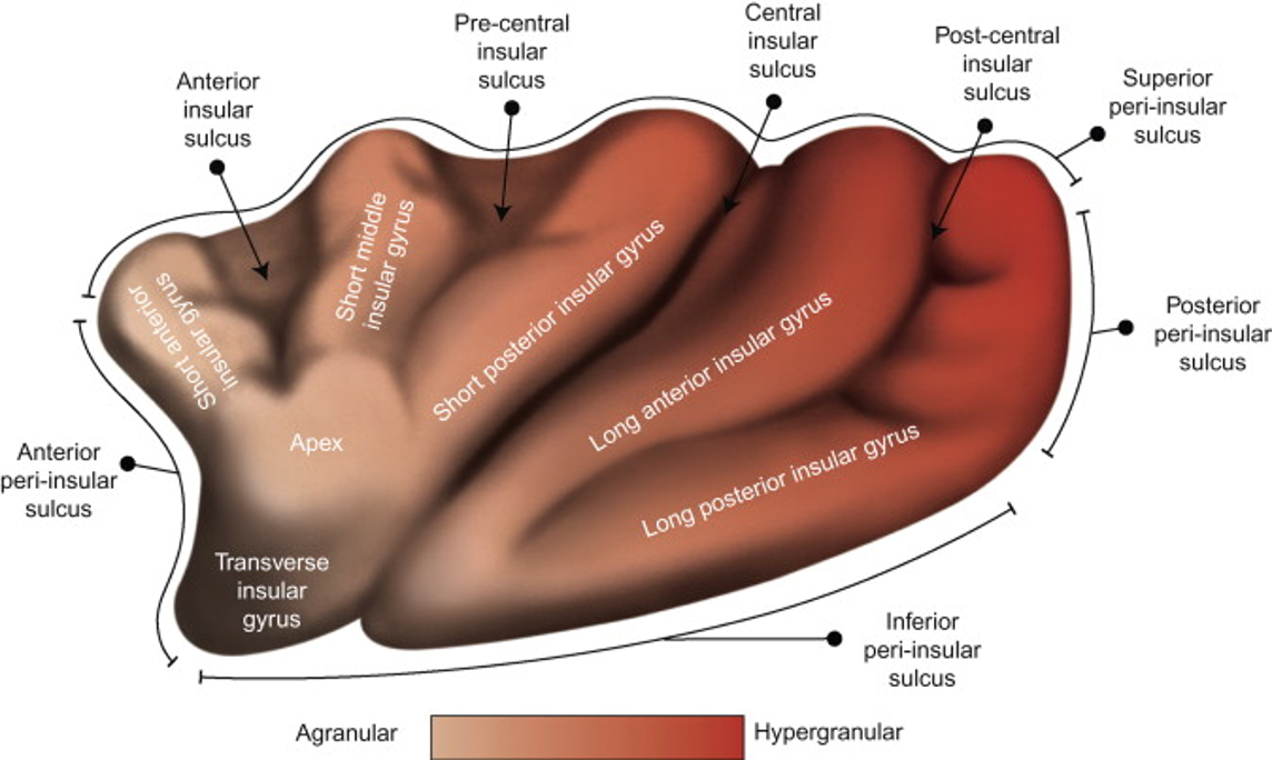
Embryological lamination of the insular cortex
- From an embryological perspective, the insular lobe is the structure between the neocortex and paleocortex.
- Three or more cytoarchitectonic cortical areas have been described in the insula, depending on the pattern of lamination. The
insular cortex is categorized based on the degree of granularity into three portions: - Central agranular cortex
- Intermediate dysgranular cortex
- The outermost granular cortex
- The level of granularity increases from the central region, that has no granular cells at all, through the intermediate that shows the presence of some granular cells, to the outermost cortex that has the fully developed granular layer.
Points
- Posterior insular point
- The point at which the superior and inferior limiting sulci meet, and
- Anterior insular point
- The point at which the anterior and superior limiting sulci meet.
Surfaces of the insula
Lateral surface is divided into two facets
- Superolateral facet
- Composed of
- Short gyri (anterior, middle, and posterior)
- Central sulcus
- Upper part of the anterior long gyrus
- Insular apex
- Edge between the anterior short gyrus and the transverse gyrus of Eberstaller that occupies the inferior portion of the anterior surface of the insula.
- Transverse insular gyrus of Eberstaller
- Is different from transverse frontal gyrus
- Is directed medially from the insular pole and is continuous with the posterior orbital gyri anteriorly.
- Relation
- Operculum of the pars triangularis, pars opercularis, and precentral and postcentral gyri.
- Superior lateral insular cleft is the space between the superolateral facet of the insula and the frontal and parietal opercula
- Inferolateral facet
- Comprised of
- Inferior portion of the anterior and posterior long gyri
- Basal continuation of the short gyri of the superolateral facet of the insula below the insular edge comprises the transverse gyrus of Eberstaller.
- The basal continuation of the short gyri courses inferomedially toward the superomedial portion of the insular pole, just lateral to the lateral olfactory striae.
- The anterior portion of the inferolateral facet is actually the base of the anterior surface of the insula.
- The posterior edge of the medial orbital gyrus and the medial edge of the posterior orbital gyrus, collectively called the posteromedial lobule, and the medial end of the transverse gyrus of the insula meet just anterior to the lateral olfactory striae, lateral to the olfactory tract (Fig. 6B).
- The inferior lateral insular cleft is the space between the inferolateral facet of the insula and the temporal operculum
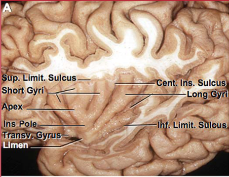
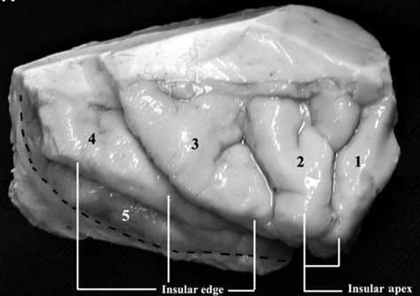
- Lateral view of the right insula
- The short insular sulcus that separates the anterior and the middle short gyri traverses the insular apex and extends to the limen insulae.
- 1, anterior short gyrus
- 2, middle short gyrus
- 3, posterior short gyrus
- 4, anterior long gyrus
- 5, posterior long gyrus.
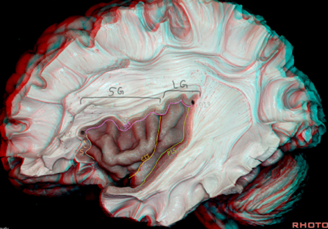
- The central sulcus of the insula is located between the posterior short gyrus and the anterior long gyrus of the insula.
- The dotted line indicates the inferior limiting sulcus of the insula.
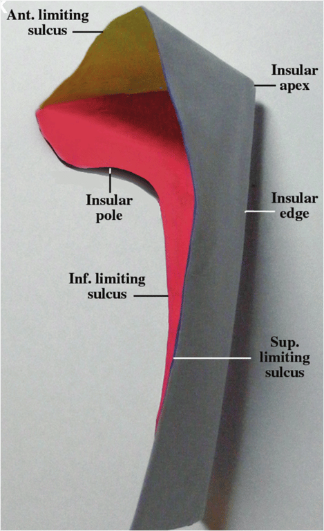
- Schematic drawing (the insula is cut out and in this picture caved in) depicting a posterosuperomedial view of the walls of the insula.
- ANT., anterior
- SUP., superior
- INS., insular
- INF., inferior
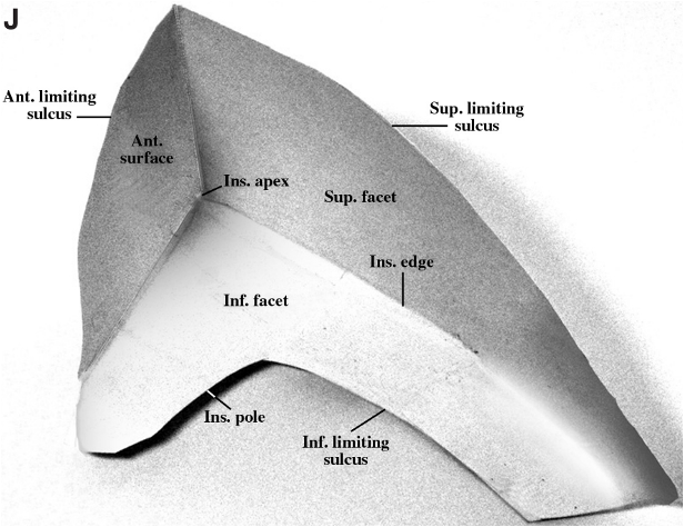
- Schematic drawing depicting a posterosuperior view of the walls of the insula.
- The anterior wall is in yellow
- The superolateral facet is in light blue
- The inferolateral facet is in red.
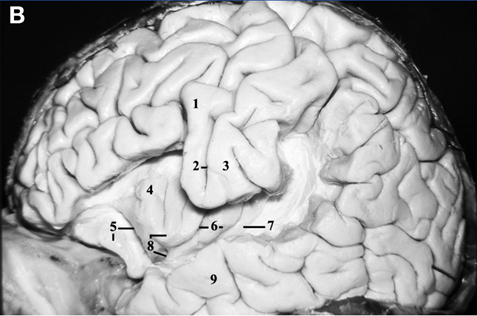
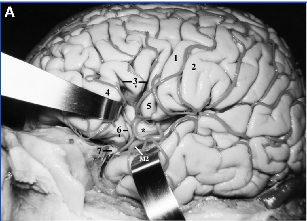
- Lateral view of the left cerebral hemisphere.
- The inferior frontal and supramarginal gyri have been removed to display the relationship between the insula and the opercula of the precentral and postcentral gyri
- 1, Precentral gyrus
- 2, Central sulcus
- 3, Postcentral Gyrus
- 4, Anterior Short gyrus of insula
- 5, Posterior orbital gyrus and the anterior insular cleft
- 6, Central sulcus and the anterior long gyrus of the insula
- 7, Posterior long gyrus of insula and inferior limiting sulcus of the insula
- 8, Insula apex and pole
- 9, Middle temporal gyrus
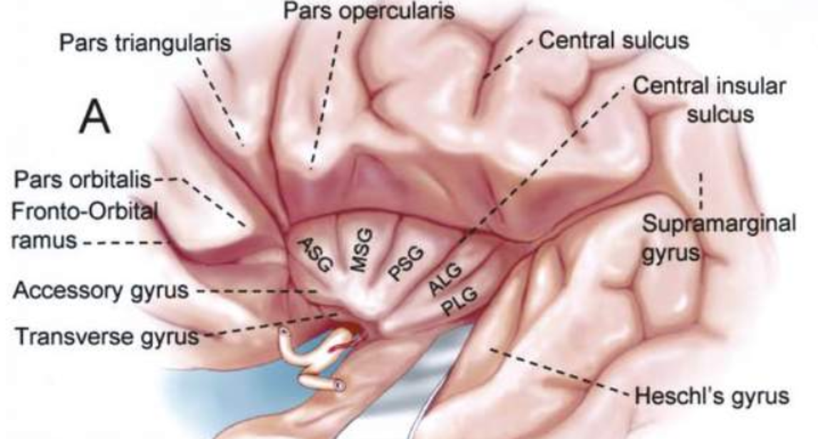
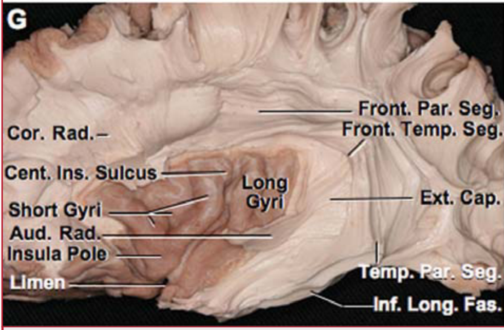
- The central insular sulcus separates the insula into larger anterior and smaller posterior portions.
- The anterior portion consists of three short gyri (anterior, middle, and posterior) arranged in a radiating pattern that converges at the insular pole located at the anteroinferior edge of the short insular gyri.
- The anterior and posterior long gyri extend backward and upward from the limen insulae.
- The distal portion of the auditory radiation courses inside Heschl’s gyrus.
- The gray matter and superficial short fibers of the inferotemporal and temporo-occipital gyrus have been removed to expose the inferior longitudinal fasciculus, which runs from the anterobasal temporal region to the occipital lobe.
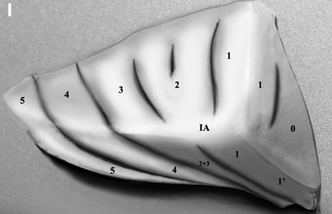
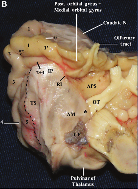
Basal surface
- Basal view of the right insula to display the inferolateral facet.
- The short insular sulcus that runs between the anterior and middle short gyri also traverses the insular apex and extends to the limen insulae.
- 0, Accessory gyrus;
- 1', Transverse insular gyrus of Eberstaller;
- 1, Anterior short gyrus;
- 2, Middle short gyrus;
- 3, Posterior short gyrus;
- 2 + 3, Fusion of the middle and posterior short gyri;
- 4, Anterior long gyrus;
- 5, Posterior long gyrus;
- IP, Insular pole;
- RI, Rhinal incisura;
- APS, Anterior perforated substance;
- TS, Temporal stem;
- OT, Optic tract;
- AM, Amygdala;
- CP, Insertion of the choroid plexus at the inferior choroidal point.
- **, Short insular sulcus.
- The dotted line indicates the inferior limiting sulcus of the insula.
- *, Medial nucleus of amygdala.
- The medial nucleus of the amygdala is located anterior and slightly superior to the inferior choroidal point, and it is in close proximity to the optic tract and the upper part of the crus cerebri.
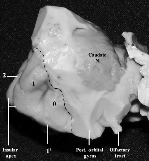
Frontal surface
- Frontal view of the right insula.
- 0, Accessory gyrus;
- 1', Transverse gyrus of Eberstaller;
- 1, Anterior short gyrus;
- 2, Middle short gyrus;
- Caudate N, head of the caudate nucleus.
- Dotted line, indicates the anterior limiting sulcus of the insula.
- The insular edge and the superolateral and inferolateral facets of the insula are more evident at the anterior portion of the insula as shown in this figure.
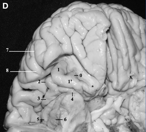
- Laterobasal view of the cerebrum.
The posterior orbital gyrus has been removed to display the overall view of the anterior surface and the inferolateral facet of the insula.
- 0, Accessory gyrus;
- 1', Transverse gyrus of Eberstaller;
- 1, Anterior short gyrus;
- 2, Middle short gyrus and the insular apex;
- 3, Posterior short gyrus;
- 4, Insular pole;
- 5, Anterior long gyrus;
- 6, Posterior long gyrus;
- 7, Horizontal ramus of the sylvian fissure;
- 8, Pars triangularis.
- *, Posteromedial orbital lobule.
- The anterior zone of the insula (anterior surface and the short gyri) forms a pyramidal structure with its apex pointing laterally and inferiorly. The insular pole and the limen insulae are located more medially and posteriorly in relation to pars triangularis
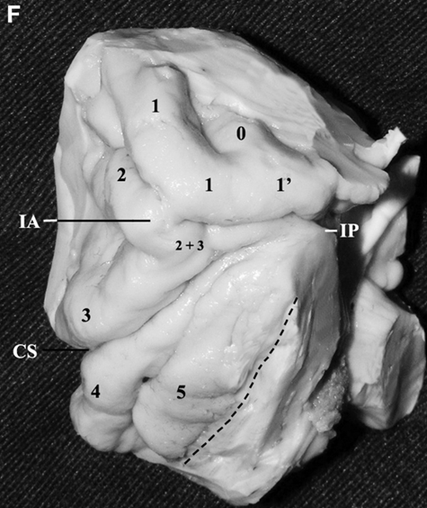
- Laterobasal view of the right insula.
- 0, Accessory gyrus;
- 1', Transverse gyrus of Eberstaller;
- 1, Anterior short gyrus;
- 2, Middle short gyrus;
- 3, Posterior short gyrus;
- 2 + 3, Junction between the middle and the posterior short gyri;
- 4, Anterior long gyrus;
- 5, Posterior long gyrus;
- IA, Insular apex;
- IP, Insular pole;
- CS, Central sulcus of insula.
- The dotted line indicates the location of the inferior limiting sulcus of insula.
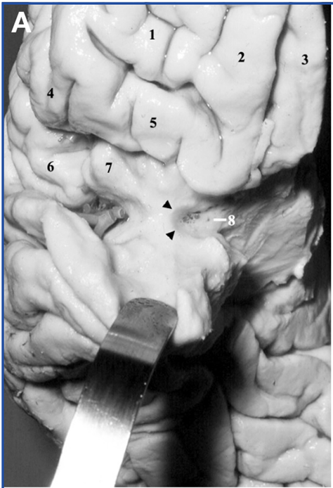
- Frontobasal view of the right hemisphere. The anterior part of the planum polare has been retracted inferiorly to display the anterior aspect of the insular pole.
- 1, Anterior orbital gyrus
- 2, Medial orbital gyrus
- 3, Rectus gyrus
- 4, Pars triangularis
- 5, Posterior orbital gyrus
- 6, Pars opercularis
- 7, Anterior short gyrus of the insula
- 8, Anterior perforating substance
- Black arrow heads, Limen insula
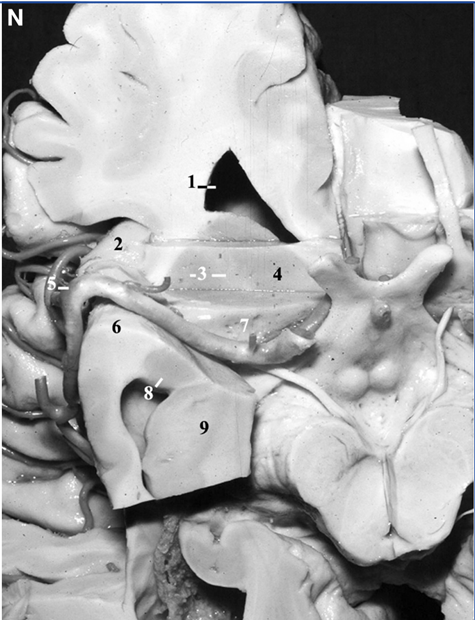
- Basal view of the right hemisphere.
- A coronal section, followed by an axial section, has been performed at the level of the anterior perforated substance.
- 1, Frontal horn;
- 2, Accessory gyrus of the insula;
- 3, Lentiform nucleus and the internal capsule;
- 4, Head of the caudate nucleus;
- 5, Genu of the MCA;
- 6, Insular pole;
- 7, Anterior perforated substance;
- 8, Amygdala;
- 9, Head of the hippocampus.
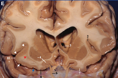
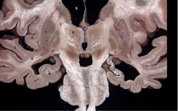
- Frontal view.
- A coronal cut has been made at the anterior portion of the insula at the level of the optic nerves. In the anterior portion of the insula, the superolateral and the inferolateral facets, as well as the insular edge, are more evident. In the right hemisphere, the coronal cut includes the extension of the anterior short gyrus in the inferolateral facet, the transverse gyrus of Eberstaller, and its junction with the posteromedial orbital lobule (blue arrow). In the left hemisphere, a coronal cut has been made more anteriorly and included the posterior orbital gyrus. At this level, the head of the caudate is located immediately above the gray matter overlying the olfactory sulcus and is separated from the latter by a thin layer of white matter. The lentiform nucleus is located above the posteromedial orbital lobule and above the transverse gyrus of Eberstaller and is separated from the latter by a layer of white matter interposed by the claustrum.
- 1, Head of the caudate nucleus;
- 2, Superior limiting sulcus;
- 3, Foramen of Monro (left);
- 4, Lentiform nucleus and olfactory sulcus;
- 5, Rectus gyrus;
- 6, Optic nerve and olfactory tract;
- 7, Posterior orbital gyrus;
- 8, Posteromedial orbital lobule.
- Arrowheads indicate superior insular cleft and superolateral facet of insula.
- White arrow indicates transverse gyrus of Eberstaller.
- Blue arrow indicates junction between the transverse gyrus of Eberstaller and the posteromedial orbital lobule.
- IE, insular edge.
- *, Paraolfactory gyrus.
- Red arrowhead indicates claustrum.
- The head and body of the caudate nucleus are located more superiorly than the superior limiting sulcus of the insula
Relations
Deep
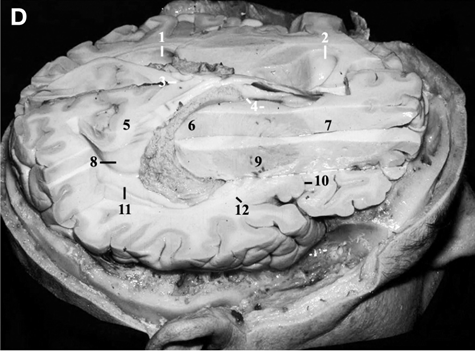
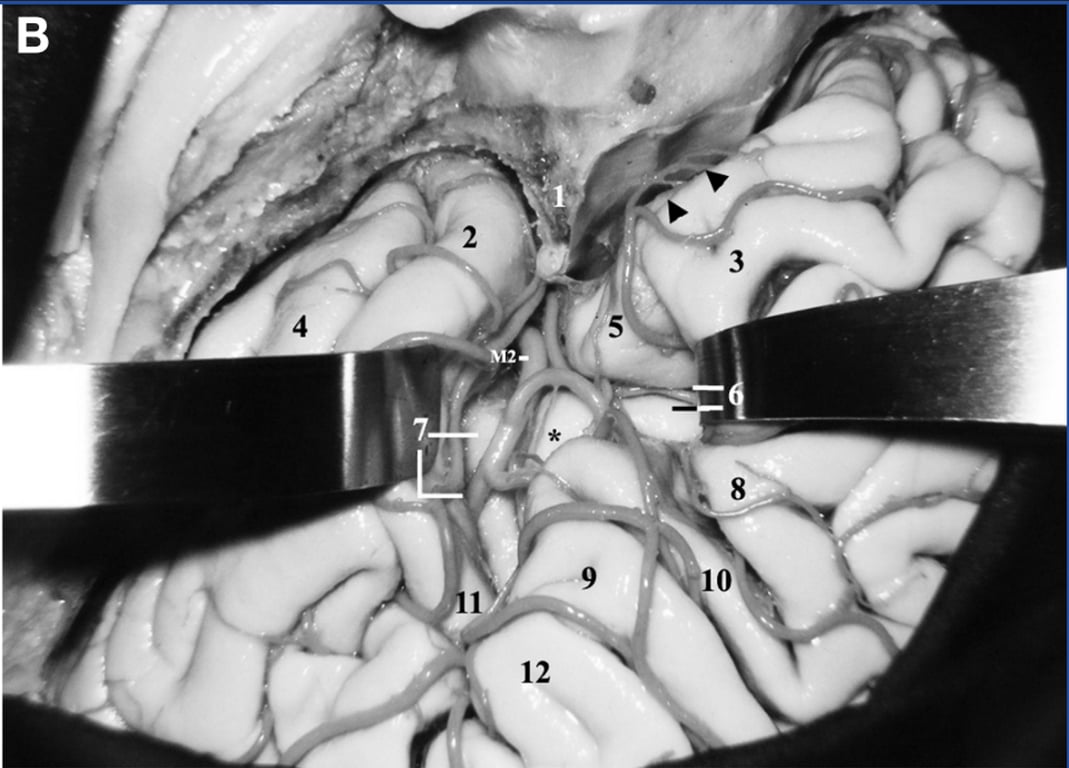
- Superolateral view of the right cerebral hemisphere.
- 1, Left atrium
- 2, Left frontal horn
- 3, Splenium of the corpus callosum;
- 4, Foramen of munro and caudate
- 5, Bulb of corpus callosum
- 6, Pulvinar
- 7, Internal capsule (anterior limb)
- 8, Calcar avis
- 9, Lentiform nucleus
- 10, Insula (anterior insular cleft)
- 11, Collateral Trigone
- 12, Sublenticular portion of the internal capsule
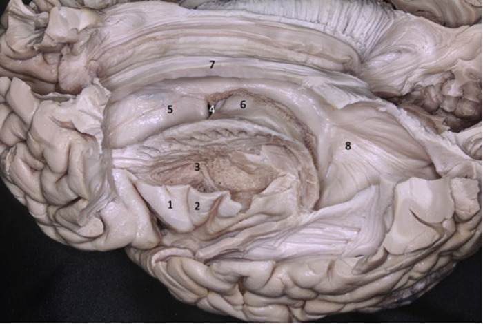
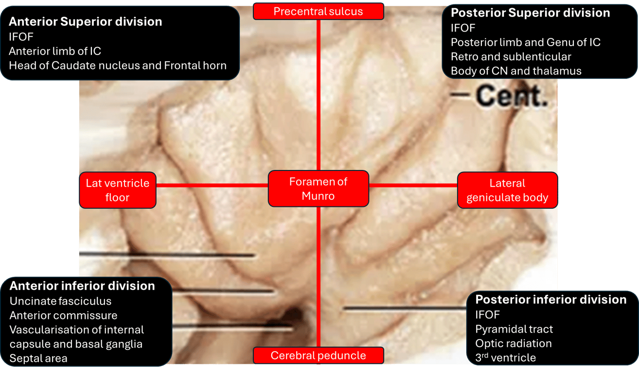
- Lateral view of the left cerebral hemisphere. Part of the frontal and temporal lobes were removed to expose the insula, basal ganglia, and ventricular system. The second short gyrus of the insula (2) aligned with the foramen of Monro (4). The foramen of Monro is located medially to the knee of the internal capsule.
- 1 - First short gyrus of the insula;
- 2 - Second short gyrus of the insula;
- 3 – Basal ganglia;
- 5 - Caudate nucleus head;
- 6 - Thalamus;
- 7 - Corpus callosum;
- 8 - Lateral ventricle atrium.
- Coronal brain section showing the relations between the insula and the gyri of the sylvian fissure. The superior temporal gyrus (1) covers the lower half of the lobe of the insula while the inferior frontal gyrus (2) covers the upper half.
- 1 - Superior temporal gyrus;
- 2 - Inferior frontal gyrus;
- 3 - Insular cortex;
- 4 - Temporal stem;
- 5 - Internal capsule;
- 6 - Thalamus;
- 7 - Lentiform nucleus;
- 8 - Frontal horn;
- 9 - Superior circular sulcus;
- 10 - Inferior circular sulcus.
Vascular relationships
- Anteroposterior (AP) view as in angiography
- 1, Parietal occipital artery
- 2, Lingual gyrus and calcarine sulcus
- 3, Calcar avis, atrium, and posterior transverse temporal gyrus
- 4, Great cerebral vein of galen
- 5, Glomus of atrium and sylvian point
- 6, Middle transverse temporal gyrus
- 7, Tentorial edge and trochlear nerve
- 8, P2P segment of the posterior cerebral artery, parahippocampal gyrus, and fornix
- 9, Sylvian point
- 10, Inferior choroidal point
- 11, Heschl’s gyrus
- 12, Lentiform nucleus
- 13, Crux cerebri
- 14, Apex and the posteromedial surface of the uncus and the anterior choroidal artery
- 15, P1 segment of the posterior cerebral artery and the posterior communicating artery
- 16, Head of hippocampus
- 17, ICA and anterior medial surface of uncus
- 18, Limen insulae and insular pole
- 19, Planum polare
- 20, Deep middle cerebral vein
- 21, Anterior cerebral artery and optic nerve;
- 22, Lesser wing of the sphenoid
- 23, Genu of the MCA
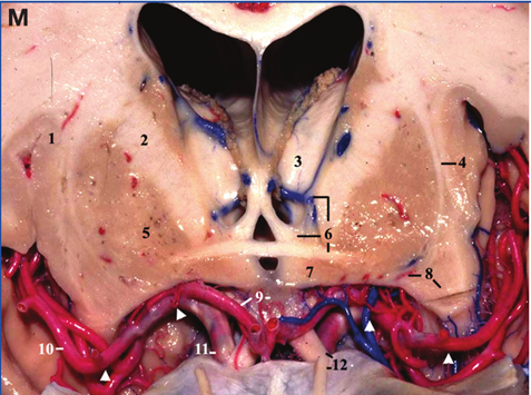
- Frontal view.
- A coronal cut has been made at the level of the bifurcation of the internal carotid artery (anterior perforated substance).
- 1, Insula;
- 2, Internal capsule;
- 3, Thalamus;
- 4, Claustrum, external and extreme capsules;
- 5, Lentiform nucleus;
- 6, Thalamostriate vein, column of the fornix, and anterior commissure;
- 7, Hypothalamus;
- 8, Lateral lenticulostriate arteries and insular pole;
- 9, Lamina terminalis and anterior cerebral artery (A1 segment);
- 10, Middle cerebral artery (MCA) (M2 segment);
- 11, Internal carotid artery (supraclinoid segment);
- 12, Olfactory tract and optic nerve.
- The arrowheads indicate the extent of the M1 segment of the MCA from the carotid bifurcation to the limen insulae.
- Superior view of the cerebrum
- 1, Orbital roof
- 2, Genu of MCA
- 3, Planum polare
- 4, Olfactory tract and optic nerve
- 5, A1 Anterior cerebral artery
- 6, Insula pole
- 7, Antero medial surface of uncus
- 8, PCOM
- 9, Hippocampus head
- 10, Anterior choroidal artery, posterior medial surface of uncus, P2A
- 11, Medial end of the Heschl’s gyrus and the sylvian point
- 12, Mid brain tegmentum
- 13, Choroid plexus (glomus) in atrium
- Lateral view of the left insula and M2 segment of the MCA
- 1, Corpus callosum
- 2, Superior limiting sulcus
- 3, Insular pole/anterior limiting sulcus
- 4, Inferior limiting sulcus
- 5, Straight sinus
- 6, Orbit
- 7, Tentorial edge and middle cranial fossa
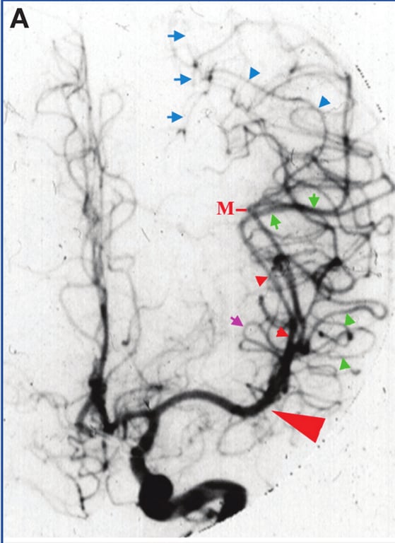
- Carotid angiography, AP view.
- Small red arrowheads: M2 segment of the MCA.
- Green arrowheads: indicate the M3 segment of the
MCA over the planum polare.
- Green arrows: M3 segment of the MCA
over the planum temporale.
- Blue arrowheads: M4 segment of the MCA
over the parietal area. - Blue arrows: the location of the intraparietal sulcus.
- Purple arrow: the superior limiting
sulcus of insula.
- Large red arrowhead: the projection of the location of the tip of the pars triangularis, just distal to the genu of the MCA (anterior sylvian point).
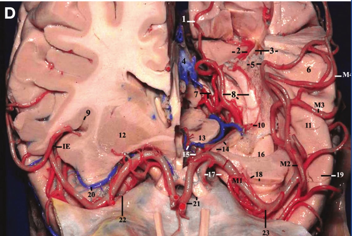
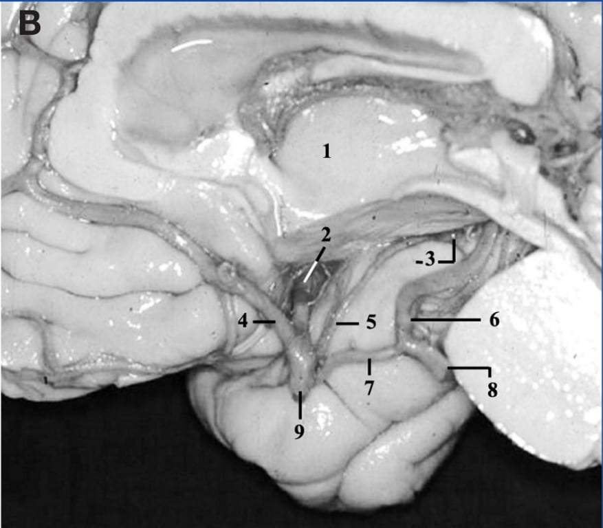
- Medial view of the right cerebral hemisphere with the crus cerebri removed to display the trajectory of the M1 segment in this projection.
- 1, Thalamus;
- 2, M1 segment of the MCA;
- 3, Uncus and the inferior choroidal point;
- 4, Anterior cerebral artery;
- 5, Anterior choroidal artery;
- 6, P2A segment of posterior cerebral artery;
- 7, Posterior communicating artery;
- 8, P1 segment of posterior cerebral artery;
- 9, Supraclinoid carotid artery.
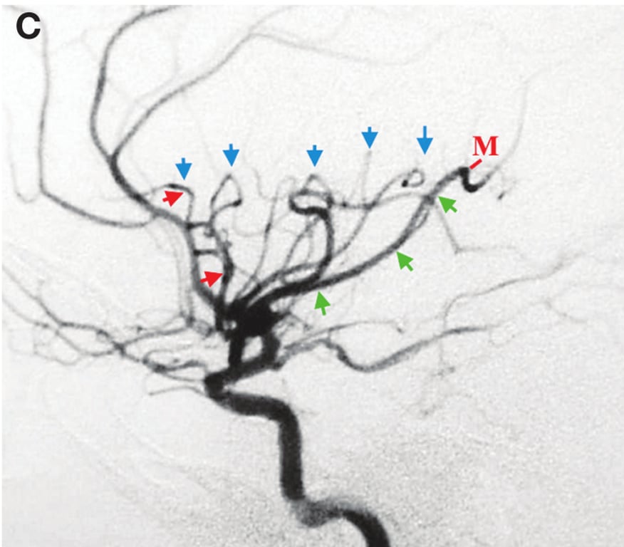
- Carotid angiography, lateral view.
- Red arrows indicate anterior limiting sulcus.
- Blue arrows indicate superior limiting sulcus.
- Green arrows indicate inferior limiting sulcus.
- M, posterior sylvian point.
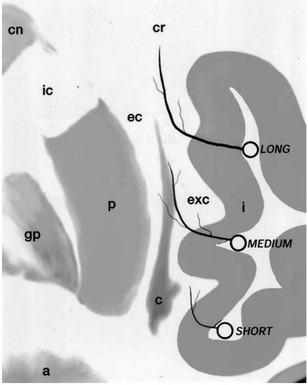
- 85 to 90% of insular arteries were short and supplied the insular cortex (i) and extreme capsule;
- 10% were medium sized and supplied, in addition, the claustrum and external capsule;
- 3 to 5% were long and extended as far as the corona radiata (cr).
- a = amygdala; gp = globus pallidus; p = putamen.
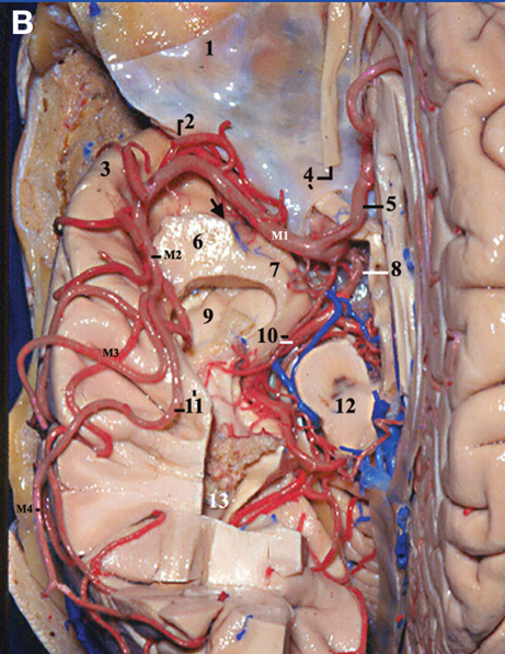
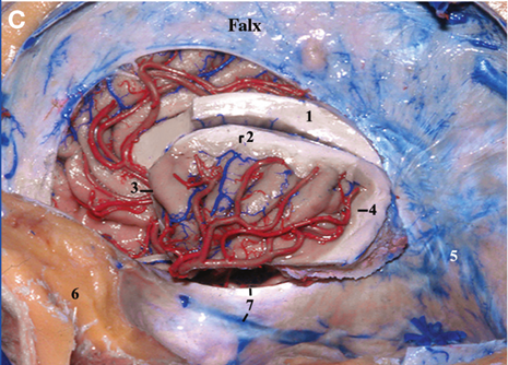
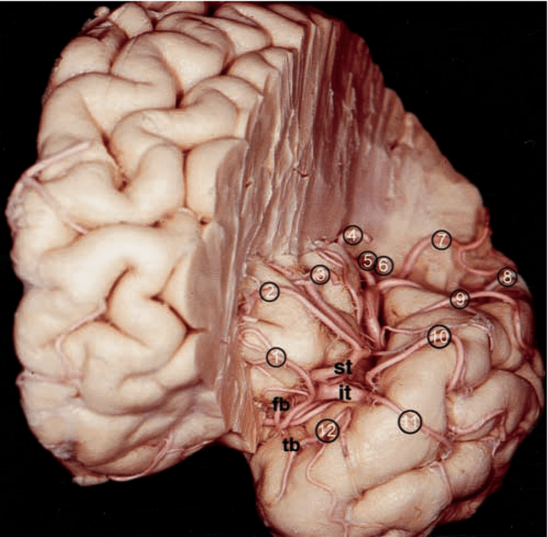
- Anterior view of the left cerebral hemisphere following removal of the frontoorbital and frontoparietal opercula. The M3 segment of the superior trunk of the MCA located in the anterior and superior periinsular sulci has been severed. The inferior trunk, which supplies the temporal lobe, is preserved.
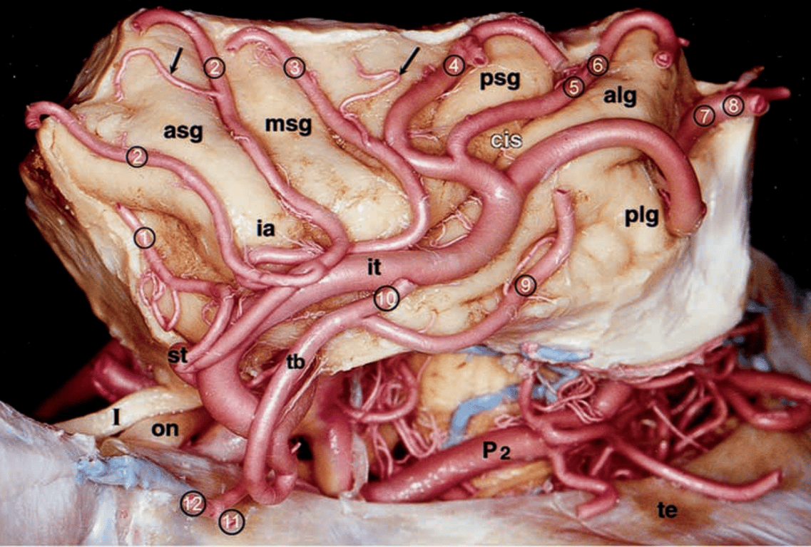
- The insula is shown following removal of the frontal, parietal, occipital, and temporal lobes from the periinsular sulci. The arteries of the insula originate from the M2 segment. The insuloopercular arteries (arrows) supply the insula and operculum.
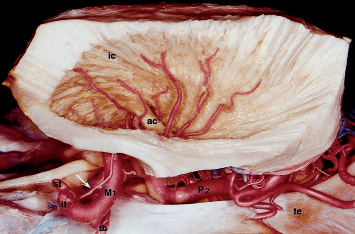
- Fiber dissection of this area reveals vascularization of the lentiform nuclei (which have been removed) and vascularization of the internal capsule by the LLAs (arrow), which arise from M1 segment.
- ac = anterior commissure; alg = anterior long insular gyrus; asg = anterior short insular gyrus; cis = central insular sulcus; ia = insular apex; ic = internal capsule; it = inferior trunk of M2; msg = middle short insular gyrus; P2 = ambient segment of the posterior cerebral artery; plg = posterior long insular gyrus; psg = posterior short insular gyrus.
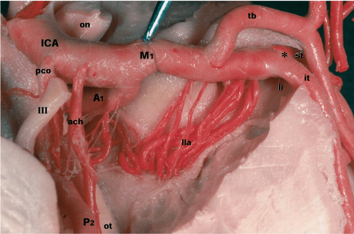
- Inferior view is shown of left MCA in the region of the anterior perforated substance following removal of the temporal lobe. The M1 segment is retracted with forceps to expose the LLAs (lla).
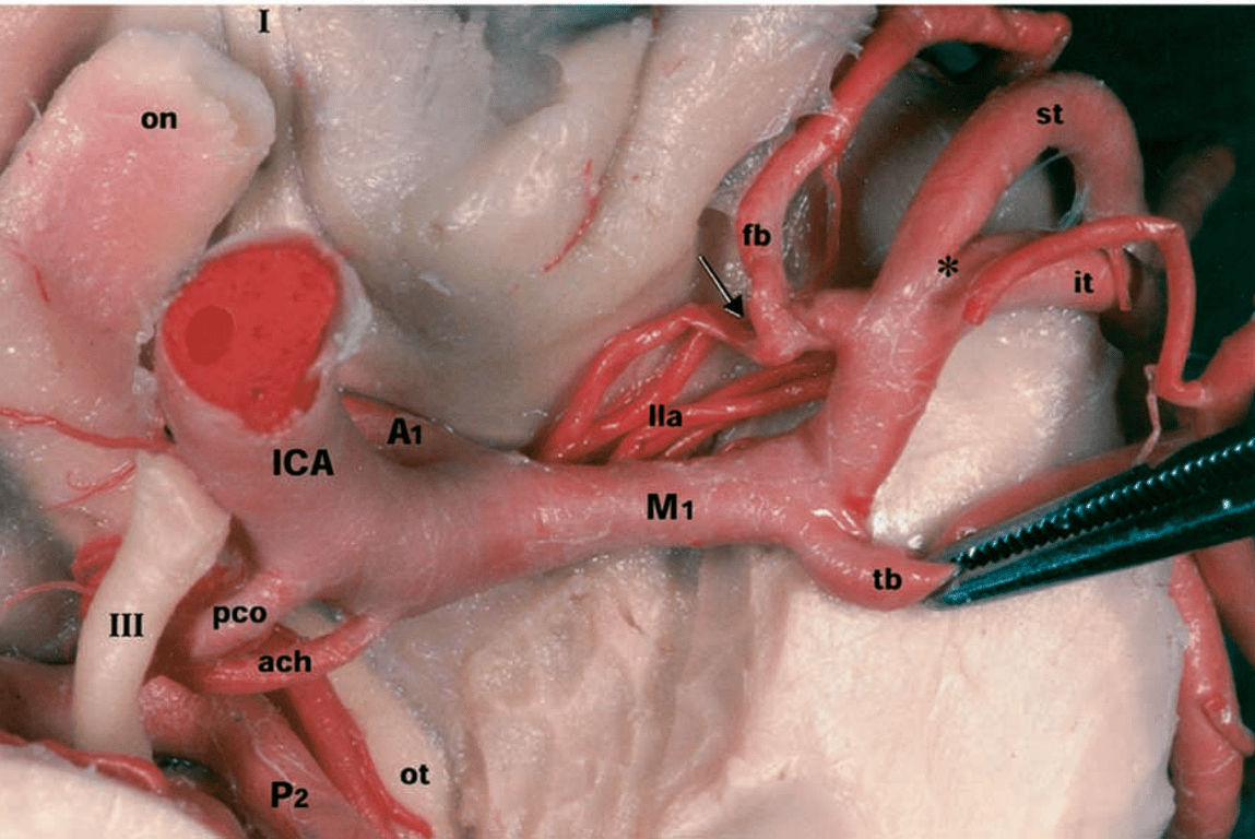
- The temporal branch of the M1 (tb) is raised to rotate the M1 segment and expose the frontal branch of the MCA (fb). The arrow indicates an LLA arising from the frontal branch (lateral orbitofrontal artery). The asterisk indicates the main bifurcation of the MCA.
- I = olfactory nerve; III = oculomotor nerve; A1 = precommunicating segment of ACA; ach = anterior choroidal artery; li = limen insulae; M1 = sphenoidal segment of the MCA; on = optic nerve; ot = optic tract; P2 = ambient segment of posterior cerebral artery; pco = posterior communicating artery.
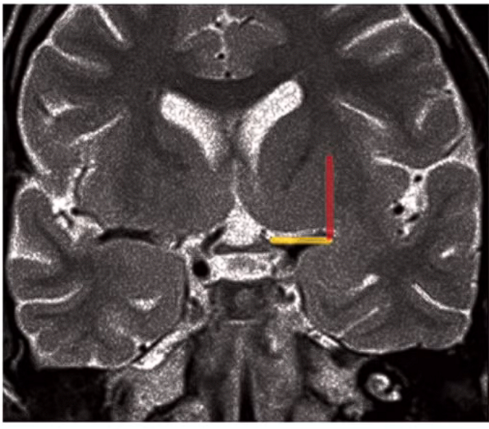
Identification of the lenticulo-striate arteries
- Coronal T2W MRI at the level of the optic chiasm
- Imaginary line drawn -from the Optic Chiasm to the Insular recess laterally — Identifies the
Anterior Perforated substance. (Yellow line)
The Portio Alegre Line (Red) runs perpendicular to the above at the lateral end and represents the lateral limit of the LSA If the medial border of the tumour crosses this line -the LSA are probably involved in the lesion
 Made with Bullet