General
- Each lateral ventricle is a C-shaped cavity that wraps around the thalamus and is situated deep within the cerebrum
Walls of lateral ventricle
Thalamus
- Located in the center of the lateral ventricle.
- Lateral ventricle wraps around the superior, inferior, and posterior surfaces of the thalamus
- Body of the lateral ventricle is above the thalamus,
- Atrium and occipital horn are posterior to the thalamus
- Temporal horn is inferolateral to the thalamus.
- The superior surface of the thalamus forms the floor of the body
- The posterior surface of the pulvinar of the thalamus forms the anterior wall of the atrium,
- The inferior surface of the thalamus is situated at the medial edge of the roof of the temporal horn.
Septum Pellucidum
- Composed of paired laminae
- Separates the frontal horns and bodies of the lateral ventricles in the midline
- Septum pellucidum attachements
- In the frontal horn, the septum pellucidum is attached to the rostrum of the corpus callosum below, the genu anteriorly, and the body above.
- In the body of the lateral ventricle, the septum is attached to the body of the corpus callosum above and the body of the fornix below.
- The septum pellucidum is tallest anteriorly and shortest posteriorly,
- Disappearing near the junction of the body and crura of the fornix where the crura and hippocampal commissure fuse with the lower surface of the corpus callosum.
- The anterior-posterior length of the septum pellucidum varies from 28 to 50 mm.
- Space between septum
- Two potential spaces between leaflets of Septum Pellucidum
- ANTERIOR: Cavum Septum Pellucidum
- A cavity with in septum pellucidum
- In the midline
- Between the laminae of the septum pellucidum.
- POSTERIOR: Cavum Vergae
- Seperation of the leaflets of septum pellucidum
- Posterior extension to the splenium of corpus collosum
- Because of ordered obliteration — Cavum Septum Pellucidium almost always accompanies a Cavum Vergae
- Obliterate Postero-anteriorly during development
- CSP present in 100% at foetal stage
- 85% Obliterate by 3-6months
- Absent CSP in fetus associated with significant CNS abnormalities
Deep cerebral white matter (Internal capsule)
- The close relationship of the internal capsule to the lateral wall of the frontal horn and body of the lateral ventricle is often forgotten in planning operative approaches to the ventricles
- The anterior limb of the internal capsule,
- Located between the caudate and lentiform nuclei
- Is separated from the frontal horn by the head of the caudate nucleus,
- The posterior limb of the internal capsule
- Located between the thalamus and the lentiform nucleus
- Is separated from the body of the lateral ventricle by the thalamus and body of the caudate nucleus.
- The genu of the internal capsule
- Comes directly to the ventricular surface and touches the wall of the lateral ventricle immediately lateral to the foramen of Monro, in the interval between the caudate nucleus and the thalamus.
2 C-shaped structures
- Fornix
- C-shaped structure that wraps around the thalamus in the wall of the ventricle
- Consists mainly of
- Hippocampomamillary tract fibers
- That originate from the hippocampus, subiculum, and dentate gyrus of the temporal lobe.
- Fimbria
- Arises in the floor of the temporal horn on the ventricular surface of the hippocampal formation and passes posteriorly to become the crus of the fornix.
- The fimbria of the fornix passes below the inferolateral part of the thalamus just lateral to the medial and lateral geniculate bodies.
- The part of the thalamus medial to the fimbria forms the roof of the ambient cistern.
- Crus of fornix
- Wraps around the posterior surface of the pulvinar of the thalamus and arches superomedially toward the lower surface of the splenium of the corpus callosum.
- At the junction of the atrium and the body of the lateral ventricle, the paired crura meet to form the body of the fornix, which runs forward along the superomedial border of the thalami in the medial wall of the body of the lateral ventricle.
- In the area below the splenium, a thin sheet of fibers interconnects the medial margins of the crura to form the hippocampal commissure.
- The crus of the fornix crosses the pulvinar approximately midway between the medial and lateral edge of the pulvinar.
- The part of the pulvinar lateral to the crus of the fornix forms part of the anterior wall of the atrium,
- The part of the pulvinar medial to the fornix forms part of the anterior wall of the quadrigeminal cistern.
- Body of the fornix
- Separates the roof of the third ventricle from the floor of the bodies of the lateral ventricles.
- At the anterior margin of the thalamus, the body of the fornix separates into two columns that arch along the superior and anterior margins of the foramen of Monro in their course toward the mamillary bodies.
- The body of the fornix crosses the thalamus approximately midway between the medial and lateral edge of the superior surface of the thalamus.
- The part of the thalamus lateral to the body of the fornix forms the floor of the body of the lateral ventricle,
- The part of the thalamus medial to the fornix forms part of the lateral wall of the velum interpositum and third ventricle.
- In the lateral ventricle's
- Body: the body of the fornix is in the lower part of the medial wall
- Atrium: the crus of the fornix is in the medial part of the anterior wall
- Temporal horn: the fimbria of the fornix is in the medial part of the floor
Images
- Septum pellucidum (orange)
- The septum pellucidum is in the medial wall of the frontal horn and body of the lateral ventricle.
- The septum pellucidum, which separates the frontal horns in the midline, does not extend to the anterior tip of the frontal horn in the lateral view because the frontal horn is directed forward and laterally from the anterior margin of the septum pellucidum.
- Thalamus (yellow): Each lateral ventricle wraps around the thalamus.
- The frontal horn is anterior to the thalamus,
- The body is above the thalamus
- The atrium and occipital horn are behind the thalamus,
- The temporal horn is below and lateral to the thalamus.
- Hippocampal formation Fornix (purple)
- The hippocampal formation is in the floor of the temporal horn.
- The fornix arises in the hippocampal formation and wraps around the thalamus in the medial part of the temporal horn, atrium, and body.
- The fimbria of the fornix arises on the surface of the hippocampal formation in the temporal horn.
- The crus of the fornix is posterior to the thalamus in the wall of the atrium.
- The body of the fornix passes above the thalamus in the lower part of the medial wall of the body.
- The columns of the fornix are formed at the level of the foramen of Monro and pass inferior to the mamillary bodies.
- The crura of the fornix are connected across the midline in the roof of the third ventricle by the hippocampal commissure.
Middle, superior view
Bottom, anterior view
- Corpus callosum (red): Made up of the
- Rostrum (which is in the floor of the frontal horn),
- Genu (which forms the anterior wall and roof of the frontal horn)
- The genu of the corpus callosum gives rise to a fiber bundle called the forceps minor, which forms the anterior wall of the frontal horn.
- Body (which forms the roof of the body of the lateral ventricle)
- Splenium
- Which gives rise to the fiber bundles making up the forceps major, which forms a prominence in the medial wall of the atrium called the bulb of the corpus callosum
- The body and splenium give rise to a fiber bundle called the tapetum, which sweeps downward to form the roof and lateral wall of the atrium and temporal horn.
- Caudate nucleus (green)
- The head and body of the caudate nucleus form the lateral wall of the frontal horn and body of the lateral ventricle.
- The tail of the caudate nucleus extends into the anterior part of the lateral wall of the atrium and into the medial part of the roof of the temporal horn to the level of the amygdaloid nucleus, which is in the anterior wall of the temporal horn.
- Fornix and hippocampal formation (purple)
- A prominence in the medial wall of the atrium, called the calcar avis, overlies the calcarine sulcus.
Middle, view through inferior surface of the hemisphere;
Bottom, view through the anterior surface of the hemisphere.
5 parts of the lateral ventricle
- The ventricular surfaces formed by the various structures are shown in different colours:
- Corpus callosum, red;
- Thalamus, yellow;
- Fornix and hippocampal formation, purple;
- Caudate nucleus, green;
- Septum pellucidum, orange;
- Prominences overlying the collateral and calcarine sulci, brown.
- 1, Splenium of the corpus callosum;
- 2, head of the caudate nucleus;
- 3, Thalamus;
- 4, genu of the internal capsule;
- 5, Anterior limb of the internal capsule;
- 6, posterior limb of the internal capsule;
- 7, Calcar avis;
- 8, glomus (choroid plexus);
- 9, Retrolentiform part of the internal capsule;
- 10, lentiform nucleus;
- 11, collateral trigone;
- 12, sublentiform part of the internal capsule and roof of the temporal horn
Frontal horn (A)
- Part of the lateral ventricle located anterior to the foramen of Monro
- Roof:
- Genu of the corpus callosum
- Anterior wall:
- Genu of the corpus callosum
- Lateral wall:
- Caudate nucleus
- Floor:
- Rostrum of corpus callosum
- Narrow floor
- Medial wall:
- Septum pellucidum
- The columns of the fornix, as they pass anterior to the foramen of Monro, are in the posteroinferior part of the medial wall.
Body of the lateral ventricle (B)
- Posterior edge of the foramen of Monro ↔ Point where septum pellucidum disappears and the corpus callosum and fornix meet
- Roof: body of the corpus callosum
- Lateral wall: Caudate nucleus
- Floor: Thalamus
- Striothalamic sulcus:
- Sulcus which separate the Caudate nucleus and thalamus
- The groove in which the stria terminalis and the thalamostriate vein course.
- Medial wall:
- Superior: Septum pellucidum
- Inferior: fornix
- The choroidal fissure
- Site of the attachment of the choroid plexus in the lateral ventricle,
- Located between the fornix and the thalamus.
Atrium (C)
- Roof: body, splenium, and tapetum of the corpus callosum
- Lateral wall:
- Anterior part: formed by the caudate nucleus as it wraps around the lateral margin of the pulvinar,
- Posterior part: formed by the fibers of the tapetum of the corpus callosum as they sweep anteroinferiorly along the lateral margin of the ventricle.
- Floor:
- Collateral trigone, a triangular area that bulges upward over the posterior end of the collateral sulcus
- Medial wall:
- The prominence that overlies the deep end of the calcarine sulcus,
- The calcarine sulcus forms a prominence, the calcar avis, in the medial wall of the atrium
- https://www.researchgate.net/figure/Horizontal-MRI-showing-the-calcarine-sulcus-a-and-the-calcar-avis-b-1-Calcarine_fig5_328941524
- Which overlies and formed by forceps major.
Inferior part: Calcar avis
The three segments of the calcarine sulcus according to Duvernoy
1, Parieto-occipital fissure; 2, Anterior part of the calcarine sulcus; 3, Calcarine sulcus proper; 4, Retro-calcarine sulcus, ascending and descending rami; 5, Temporo-occipital fissure; 6, Lingual sulcus; 7, Collateral sulcus (medial occipitotemporal sulcus); O3, Third occipital gyrus; O4, Fourth occipital gyrus; O5, Fifth occipital gyrus (Lingual gyrus); O6, Sixth occipital gyrus (Cuneus)
Number of peaks along the calcarine sulcus course. A, One peak (white arrow); B, three peaks (white arrowheads).
1, Parieto-occipital fissure; 2, Calcarine sulcus proper; 3, Retrocalcarine sulcus with its ascending and descending rami; 4, Paracalcarine sulcus
A, Discontinuous character of the calcarine sulcus; B, collateral branches along its course (white arrowheads).
1, Parieto-occipital fissure; 2, Anterior part of the calcarine sulcus; 3, Calcarine sulcus proper
Coronal MRI passing through the anterior part of the calcarine sulcus (A) and the calcar avis (B).
1, Anterior part of the calcarine sulcus; 2, Occipital horn of the lateral ventricle; 3, Calcar avis
Horizontal MRI showing the calcarine sulcus (A) and the calcar avis (B).
1, Calcarine sulcus; 2, Occipital horn of the lateral ventricle; 3, Calcar avis
Superior part: bulb of the corpus callosum,
- Anterior wall
- Medial part: crus of the fornix as it wraps around the posterior part of the pulvinar,
- Lateral part: pulvinar of the thalamus.
- In the atrium, the choroid plexus has a prominent tuft called the glomus.
Occipital horn
- Extends posteriorly into the occipital lobe from the atrium
- It varies in size from being absent to extending far posteriorly in the occipital lobe, and it may vary in size from side to side.
- Medial wall:
- Bulb of the corpus callosum + calcar avis
- Roof:
- Tapetum
- Lateral wall:
- Tapetum
- Floor:
- Collateral trigone
Temporal horn (D)
- Extends forward from the atrium below the pulvinar into the medial part of the temporal lobe and ends blindly in an anterior wall that is situated immediately behind the amygdaloid nucleus
- Roof:
- Medial part:
- Inferior surface of the thalamus
- Tail of the caudate nucleus
- Which are separated by the striothalamic sulcus.
- Lateral part
- Tapetum of the corpus callosum,
- Which also sweeps inferiorly to form the lateral wall of the temporal horn.
- Tapetum separates the temporal horn from the optic radiations.
- Lateral wall
- Tapetum of the corpus callosum,
- Floor:
- Medial part: prominence overlying the hippocampal formation,
- Lateral part: prominence called the collateral eminence, which overlies the deep end of the collateral sulcus that separates the parahippocampal and occipitotemporal gyri on the inferior surface of the temporal lobe.
- Medial wall:
- The only structure in the medial wall is the narrow cleft, the choroidal fissure, situated between the inferolateral part of the thalamus and the fimbria of the fornix.
Choroidal Fissure and Choroid Plexus
Choroid plexus
- The choroid plexus does not extend into the frontal horn or the occipital horn.
- The choroid plexus from each lateral ventricle extends through the foramen of Monro and is continuous with the two parallel strands of choroid plexus in the roof of the third ventricle.
- In the atrium, the choroid plexus forms a prominent triangular tuft called the glomus.
- The edges of the thalamus and fornix bordering this choroidal fissure have small ridges, called the teniae, along which the tela choroidea, the membrane in which the choroid plexus arises, is attached.
- The tenia on the thalamic side is called the tenia thalami or tenia choroidea.
- The tenia on the forniceal side of the fissure is called the tenia fornicis, except in the temporal horn where it is referred to as the tenia fimbriae.
Vascular supply
Arterial Supply of the Choroid Plexus
- The choroidal arteries converge on and pass through the choroidal fissure, which is the cleft between the fornix and the thalamus.
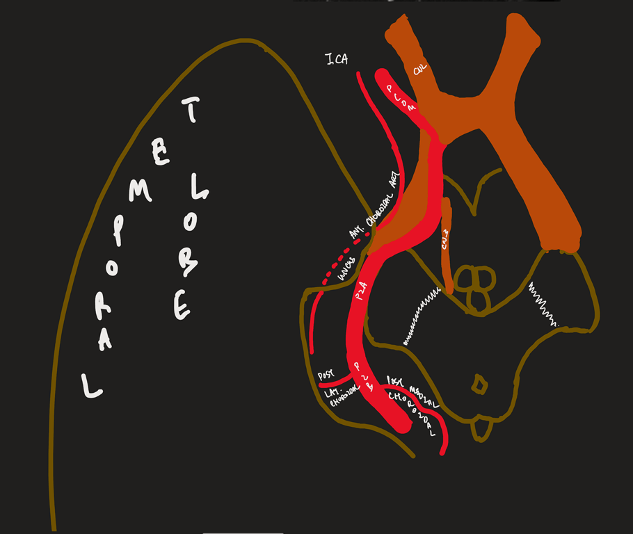
Anterior choroidal artery (ACh)
- Origin
- (1–3 mm distant to the PComA origin) usually arises from the posterior aspect of the ICA
- Course:
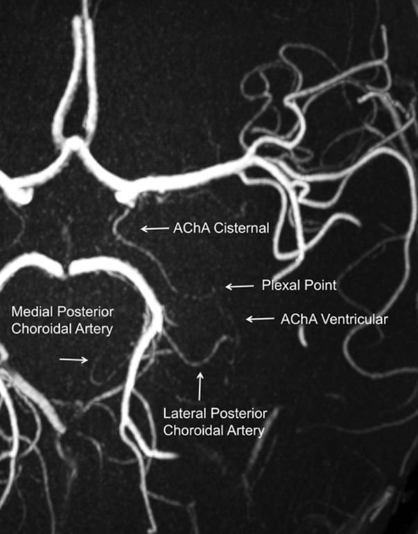
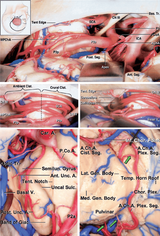
- Lateral to optic tract, curves medially to be inferomedial to optic tract --> curve laterally to run along the lateral aspect of the optic tract --> circumvents the cerebral peduncles to reach the lateral geniculate body --> traverses in the posterolateral direction above the uncus to enter the choroidal fissure, at the plexal point.
- Plexal point is always constant
- Two segments
- Cisternal segment:
- Extends from its origin until the choroidal fissure; measures ~2.5 cm
- Plexal/Intraventricular segment (3-10 perforators)
- Plexal point: where anterior choroidal artery enter temporal horn
- Supplies
- Visual system: Inferior optic chiasm, Posterior portion of optic tract, Optic radiation, Lateral geniculate body
- Temporal lobe: Uncus, Parahippocampal gyrus, Amygdala, Choroid plexus, Temporal horn, Atrium
- Basal ganglia: Globus pallidus medial, Tail of caudate, Internal capsule (genu)
- Diencephalon: Subthalamus, Thalamus (Lateral ventrolateral nucleus, Lateral ventroanterior nucleus)
- Midbrain: Middle 1/3 of cerebral peduncle, Upper red nucleus, Substantia nigra
- Very rare to have perforating branches outside of origin of anterior choroidal artery
- Anastomoses with lateral posterior choroidal artery
- Anterior choroidal artery-Clinical
- Use to be ligated to tx Parkinson's in the past
- Aneurysm: located superior/superiorlaterally to origin of anterior choroidal artery
- Stroke → Anterior choroidal artery syndrome: (3H)
- Hemisensory loss
- Hemiplegia
- Homonymous hemianopia
Difference between PCOM vs Anterior choroidal artery
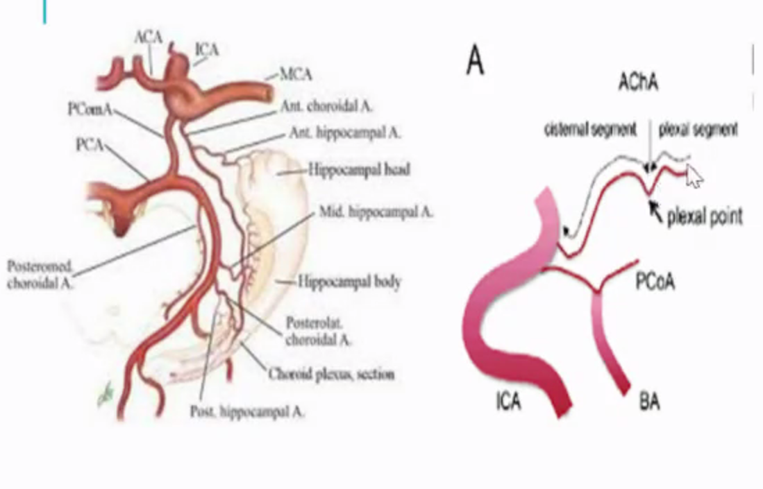
Features | Anterior choroidal | PComA |
Origin | More distal | More proximal |
Size | Smaller | Larger |
Direction of travel | Has a superior hump (plexal point) where it passes through the inferior choroidal point to enter the temporal horn. | Goes up and down the straight back up and usually bifurcates |
Relation between each | More lateral | More medial |
Posterior Choroidal Arteries (PChAs)
- Branches of the posterior cerebral artery (PCA) and are divided into two groups:
Lateral posterior choroidal artery (LPChAs)
- Origin: They arise from the PCA (most commonly the P2P segment) or its cortical branches in the ambient and quadrigeminal cisterns.
- Course
- They pass laterally around the pulvinar
- Course laterally along the upper edge of the parahippocampal gyrus within the ambient cistern
- Pass through the choroidal fissure to enter the posterior part of the temporal horn and atrium
- enters the ventricle adjacent to the lateral geniculate nucleus through the choroid fissure
- Destination: temporal horn of lateral ventricle
- The number of LPChAs in a hemisphere averages four (ranging from one to nine).
- Supplies:
- Cerebral peduncle
- Posterior commissure
- Fornix (Part of crura and body)
- Lateral geniculate body
- Pulvinar
- Dorsomedial thalamic nucleus
- Body of the caudate nucleus
- Anastomosis with
- Ant Choroidal Artery
- Medial Posterior Choroidal Artery
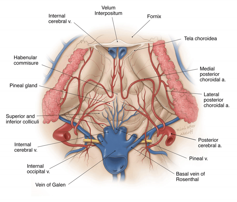
Medial posterior choroidal artery (MPChAs)
- Origin:
- They arise most frequently from the posteromedial aspect of the proximal part of the PCA (P1 or P2) in the interpeduncular and crural cisterns.
- Course
- Travels inferior and medial to the PCA through the crural → ambient cisterns and turns medially to enter the quadrigeminal cistern.
- Passes beneath the splenium of the corpus callosum
- The artery then turns forward to enter the velum interpositum and supplies the choroid plexus in the roof of the third ventricle
- Destination: Lateral + 3rd ventricle
- Supplies:
- Cerebral peduncle
- Tegmentum
- Geniculate bodies (medial > lateral)
- Colliculi
- Pulvinar
- Pineal gland
- Medial thalamus
Arterial Supply of the Ventricular Walls
The walls of the lateral ventricle (formed by structures such as the caudate nucleus, internal capsule, thalamus, and corpus callosum) are supplied by perforating arteries that arise from the major cerebral trunks in the basal cisterns:
- Internal Carotid/Anterior Choroidal Artery (AChA):
- The AChA sends branches (other than those supplying the plexus) to deep structures near the ventricular walls, including the genu and posterior limb of the internal capsule, the adjacent part of the globus pallidus, and the thalamus.
- Anterior Cerebral Artery (ACA):
- The pericallosal branches penetrate the corpus callosum and reach the septum pellucidum and the fornix.
- These are crucial components of the medial wall of the frontal horn and body.
- Middle Cerebral Artery (MCA):
- The lenticulostriate arteries arise from the MCA and supply structures lateral to the frontal horn and body, including the putamen, the entire length of the internal capsule, and the body and head of the caudate nucleus.
- Posterior Cerebral Artery (PCA) Perforators:
- Both LPChAs and MPChAs send branches along their course to adjacent neural structures.
- LPChAs may send branches to the pulvinar, lateral geniculate body, and body of the caudate nucleus.
- MPChAs may send branches to the pulvinar and medial thalamus.
Venous drainage
- Veins are coursing in the walls of the ventricles
- Exit the ventricles by passing through the margin of the choroidal fissure in the subependymal location to reach the internal cerebral, basal, or great veins.
Ventricular Part | Medial Group Veins (Drains medial wall/roof) | Lateral Group Veins (Drains lateral wall/floor) | Primary Terminating Trunk |
Frontal Horn | Anterior Septal Veins | Anterior Caudate Veins | Internal Cerebral Vein (ICV) |
Body | Posterior Septal Veins | Thalamostriate Vein (most well-known), Thalamocaudate Vein, Posterior Caudate Veins | Internal Cerebral Vein (ICV) (via velum interpositum) |
Atrium | Medial Atrial Veins | Lateral Atrial Veins | Basal, Internal Cerebral, or Great Vein of Galen (in the quadrigeminal cistern) |
Temporal Horn | Transverse Hippocampal Veins (drains floor) | Inferior Ventricular Vein (drains roof) | Basal Vein (of Rosenthal) (in crural/ambient cistern |
Surgery
- Opening through the fissure from the lateral ventricle during intracranial operations provides access to several structures that are difficult or impossible to expose through the extracerebral route.
- The velum interpositum, through which the internal cerebral veins course, is located on the medial side of the body portion of the fissure in the roof of the third ventricle.
- Opening through the choroidal fissure from the body of the ventricle will expose the velum interpositum and the roof of the third ventricle.
- Some operative approaches to the superior part of the choroidal fissure are directed through the frontal horn and adjacent part of the body.
- The atrial part is located in the atrium of the lateral ventricle between the crus of the fornix and the pulvinar (Figs. 5.5 and 5.8).
- The quadrigeminal cistern, the pineal region, and the posterior portion of the ambient cistern can be exposed by opening through the fissure from the atrium.
- The temporal part is situated in the temporal horn between the fimbria of the fornix and the inferolateral surface of the thalamus
- Opening through the choroidal fissure in the temporal horn exposes the structures in the ambient and posterior part of the crural cisterns.
- The cisternal side of the temporal portion of the fissure is situated in the superolateral edge of the ambient cistern.
Stepwise dissection from the Lateral ventricle → 3rd → Choroidal fissure →Basal cistern
A., artery; Ant., anterior; Bas., basilar; Call., callosum; Caud., caudate; Cer., cerebral; Ch., choroidal; Chiasm., chiasmatic; Chor., choroid; Col., column; Coll., colliculus; Comm., commissure; Corp., corpus; CN, cranial nerve; For., foramen; Front., frontal; Gen., geniculate; Infund., infundibular; Int., intermedia, internal; Lam., lamina; Lat., lateral; Mam., mamillary; M.P.Ch.A., medial posterior choroidal artery; Nucl., nucleus; P.C.A., posterior cerebral artery; Pell., pellucidum; Plex., plexus; Rec., recess; Sept., septal, septum; Tent., tentorial; Term., terminalis; Thal. Str., thalamostriate; V., vein; Vent., ventricle
- Superior view of the lateral ventricles.
- The choroidal fissure is the cleft between the fornix and the thalamus along which the choroid plexus is attached. The frontal horn is located anterior and the ventricular body behind the foramen of Monro.
- The thalamus forms the floor of the body of the lateral ventricle and the anterior wall of the atrium
- Enlarged view.
- The columns of the fornix form the anterior and superior margins of the foramen of Monro.
- The choroid plexus in the body extends through the posterior margin of the foramen of Monro and is continuous with the choroid plexus in the roof of the third ventricle.
- The right thalamostriate vein passes through the posterior edge of the foramen of Monro and the left thalamostriate vein passes through the choroidal fissure behind the foramen.
- Floor of the frontal horn rostrum of the corpus callosum
- Anterior wall is formed by the genu of the corpus callosum.
- Lateral wall is formed by the caudate nucleus.
- The septum pellucidum is attached to the upper edge of the body of the fornix.
- Enlarged view of the right foramen of Monro.
- The columns of the fornix form the anterior and superior margins of the foramen.
- An anterior septal vein passes backward along the septum pellucidum and crosses the column of the fornix.
- The thalamostriate vein passes forward between the caudate nucleus and thalamus and turns medially to pass through the posterior margin of the foramen of Monro to empty into the internal cerebral vein.
- The choroid plexus is attached medially by the tenia fornix to the body of the fornix and laterally by the tenia thalami to the thalamus.
- The transchoroidal exposure is begun by dividing the tenia fornix that attaches the choroid plexus to the margin of the fornix.
- The tenia thalami that attaches the choroid plexus to the thalamus is not opened.
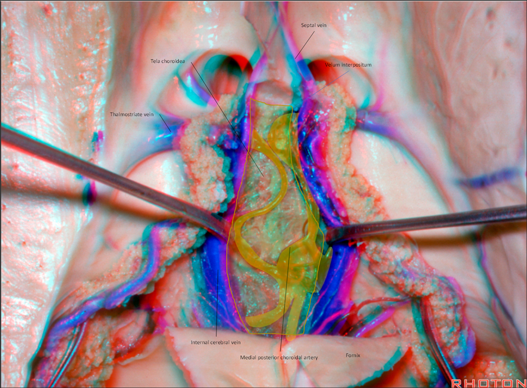
- The opening of the choroidal fissure has been extended backward from the foramen of Monro to expose both internal cerebral veins and the medial posterior choroidal arteries coursing in the velum interpositum.
- The anterior septal vein crosses the septum pellucidum.
- The lower layer of tela choroidea, attached to the striae medullaris thalami deep to the internal cerebral veins, is intact.
The lower layer of tela choroidea that forms the floor of the velum interpositum has been opened, exposing the massa intermedia and posterior commissure within the third ventricle.
The internal cerebral veins have been separated to expose the anteroinferior part of the third ventricle.
The upper end of the midbrain forms the posterior part of the floor of the third ventricle
The mamillary bodies are situated in the midportion of the floor.
The floor anterior to the mamillary bodies and behind the infundibular recess in very thin and is the site commonly opened in a third ventriculostomy.
The chiasmatic recess extends forward above the posterior edge of the optic chiasm and below the anterior commissure
Enlarged view of the inner surface of the anterior wall of the third ventricle.
The columns of the fornix extend downward behind the anterior commissure toward the mamillary bodies.
The lamina terminalis, chiasmatic recess, posterior edge of the chiasm, and the infundibular recess are located along the anterior and lower wall of the third ventricle
The opening along the choroidal fissure has been extended posteriorly by opening the tenia fornix along the edge of the body and crus of the fornix.
The upper part of the quadrigeminal cistern, where the internal cerebral veins converge on the vein of Galen, has been exposed.
The medial posterior choroidal arteries course with the internal cerebral veins
The opening of the choroidal fissure has been extended downward along the choroidal fissure to the central part of the quadrigeminal cistern, exposing the basal and internal cerebral veins, pineal, and superior colliculus. Branches of the medial posterior choroidal arteries course beside the pineal.
Enlarged view. The tip of the pineal projects posteriorly above the superior colliculus and between the terminal part of the internal cerebral veins.
The dissection has been extended forward along the choroidal fissure toward the temporal horn by dividing the tenia on the edge of the fimbria of the fornix to expose the basal vein, posterior cerebral arteries, and trochlear nerve in the posterior part of the ambient cistern below the thalamus
The choroidal fissure in the temporal horn has been opened by dividing the tenia fimbria. The choroid plexus attachment to the thalamus has not been disturbed. The posterior cerebral artery and basal vein course through the ambient cistern on the medial side of the temporal portion of the choroidal fissure.
The exposure has been extended through the amygdala anterior to the choroidal fissure to expose the oculomotor nerve and origin of the posterior cerebral artery. The posterior cerebral artery passes above the oculomotor nerve.
 Made with Bullet