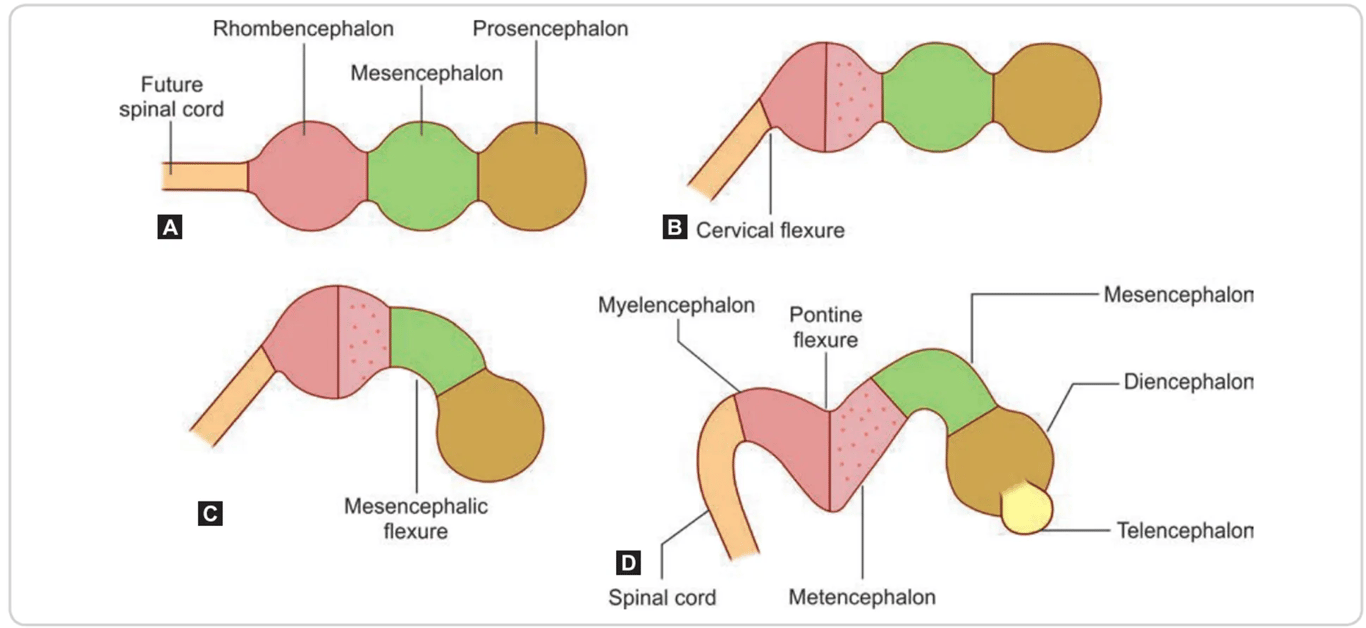
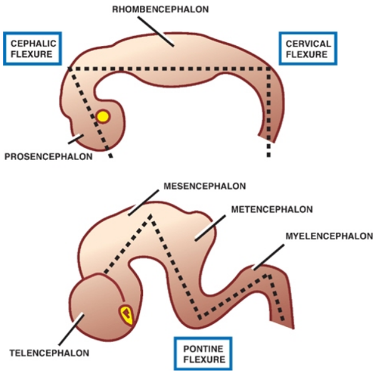
- As the primary brain vesicles develop, the brain flexes (bends) to form:
- Mesencephalic flexure (or cephalic flexure)
- In the region of the midbrain
- The mesencephalon and the rhombencephalon are separated by a constriction called the isthmus rhombencephalii
- Cervical flexure:
- Junction of the hindbrain and the spinal cord
- Compensatory pontine flexure:
- Forms between the cephalic and cervical flexures.
- At the middle of the rhombencephalon,
- Dividing it into the metencephalon and myelencephalon
- Bending at pontine flexure causes the 4th ventricle to look like a look like a rhomboid shape.
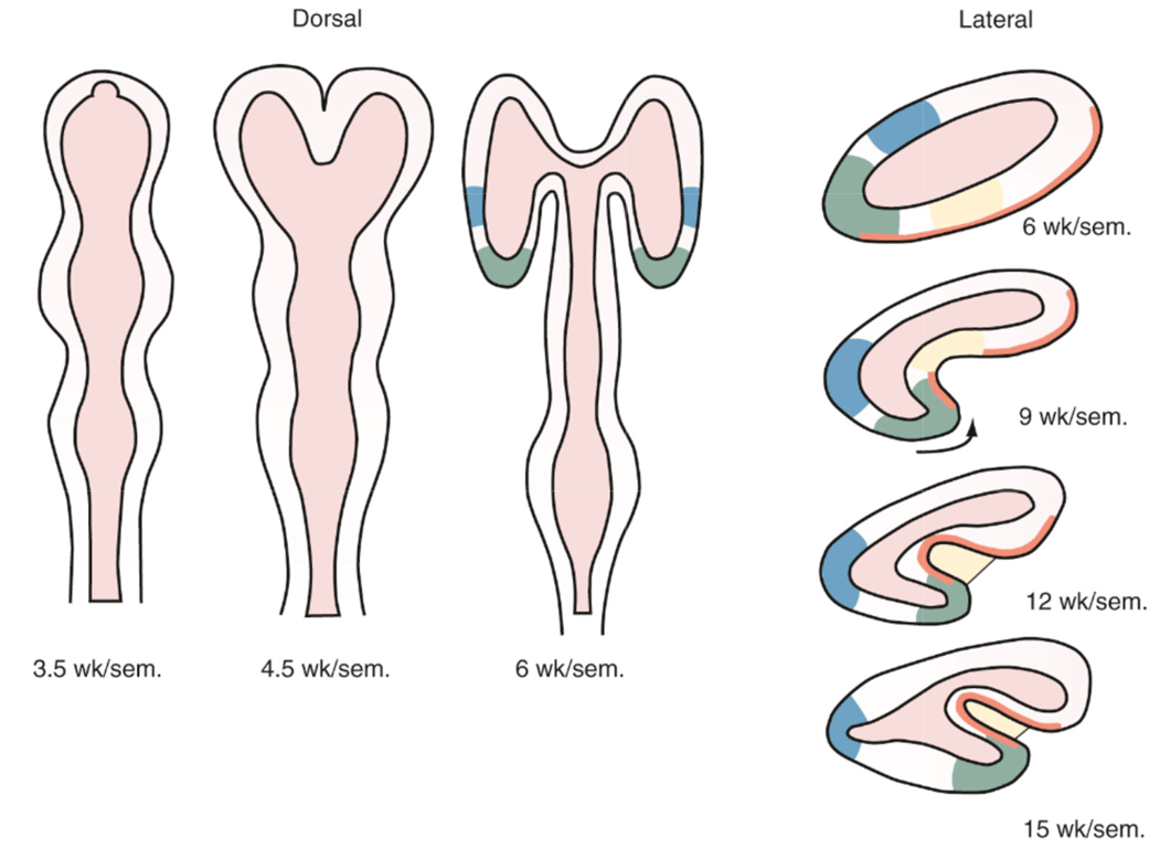
- Telencephalic flexure
- Forms the sylvian fissure
- both the dorsal (frontal) and ventral (temporal) lips of the fissure are derived from the ventral part of the primitive telencephalon (ORANGE)
- Ventrodorsal genetic gradients in the vertical axis can affect both lips, as in schizencephaly.
- The insula is an infolding of tissue secondary to bending of the hemisphere.
- Original posterior pole of the primitive telencephalic hemisphere becomes the temporal, not the occipital pole of the mature brain.
- The occipital horn of the lateral ventricle is a new recess that forms after folding of the telencephalon.
 Made with Bullet