Prosencephalon (Forebrain)
- 32 day: horizontal cleavage
- Germinal matrix begins to cleave into superior and inferior portions.
- Superior portion → telencephalon (the caudate, putamen, and cerebral hemispheres)
- Inferior portion → diencephalon (the thalamus, hypothalamus, and Globus pallidus).
- Optic vesicles
- Formed early in development
- A lateral outgrowth on each side of the forebrain.
- Give rise to the origin to the retinas and optic nerves
- Divide the forebrain into
- Telencephalon (rostral)
- Diencephalon (caudal)
- Optic vesicles themselves are of diencephalic origin
- Further differentiation of the prosencephalon into
- Most extensive developmental changes in the nervous system.
- Cerebral hemispheres
- Cerebral commissures
- Corpus striatum,
- Internal capsule.
- Early in development, the telencephalon consists of a
- Filled by the rostral extension of the third ventricle.
- Develops into the cerebral hemispheres
- Starts at 5th gestational week
- 35 day: vertical cleavage (see horizontal cleavage)
- Evagination and separation of the cerebral hemispheres
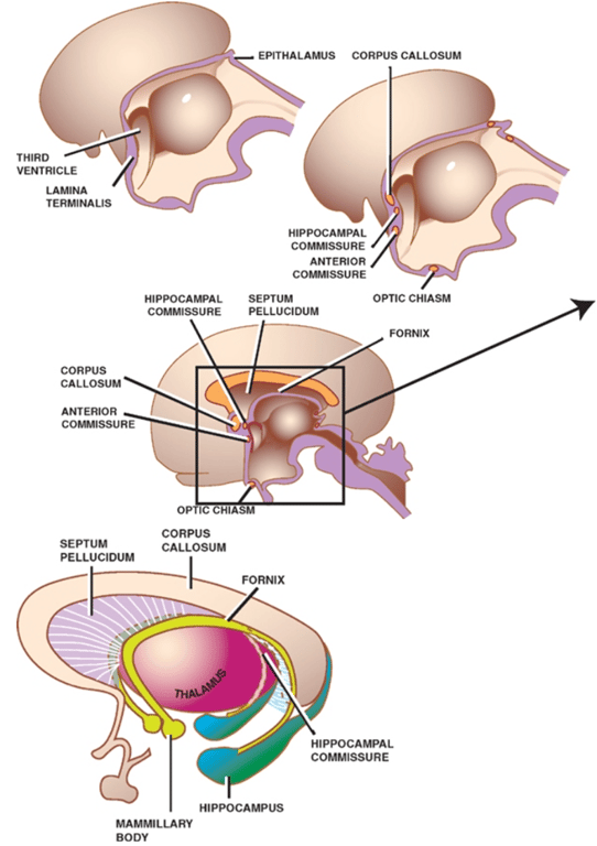
- 32-34 day lamina terminalis begins to differentiate into the inter-hemispheric cerebral commissures
- Groups of fibres that connect regions of the two cerebral hemispheres
- Originally this done by the lamina terminalis
- Forms the anterior wall of the 3rd ventricle
- Eventually 3 major commissures develop within the lamina terminalis
- 7th wk
- First commissure to form.
- Connects the olfactory bulbs and temporal lobes of both sides.
- 9th wk
- Between fornix is this hyperlink correct????
- White matter fibres between hippocampus to mamillary bodies
- Largest of the cerebral commissures, takes the form of an arch over the third ventricle.
- Connects the neocortices of both sides.
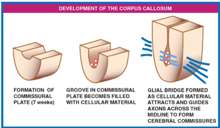
- Development
- begins at 9th week of gestation
- Dorsal aspect of the lamina terminalis thickens (commissural plate)
- A groove develops in the commissural plate,
- Cellular material fills this groove.
- Cellular material forms a glial bridge superiorly across the groove, the cellular components of which express surface molecules and secrete chemical messengers that attract and help guide axons across the midline to form the three cerebral commissures.
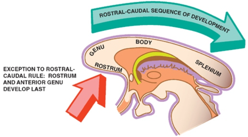
- Development of the entire corpus callosum, however, does not occur simultaneously; rather, it follows a rostral to caudal sequence.
- Arrest of the corpus callosum development prior to its completion results in a normally formed anterior portion but an absent or only partially formed posterior portion.
- Exceptions: leads to absent or small genu or body and an intact splenium and rostrum.
- Rostral most portions of the corpus callosum—the rostrum and the anterior part of the genu.
- Which form a bit later
- Secondary destructive processes that damage the corpus callosum after it has already fully formed.
- Semilobar Holoprosencephaly
- Septum pellucidum
- What is left of the lamina terminalis after the development of the 3 commissures is a thin wall
- This separates the anterior horns of the lateral ventricles.
- The corpus callosum and the fornix bound the anterior horns of the lateral ventricles from above and below, respectively.
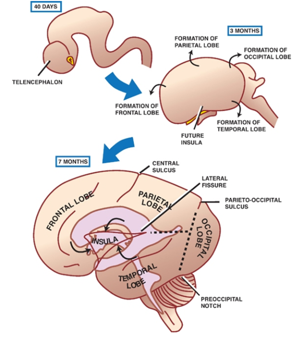
- Falx cerebri:
- Formed from a trapped embryonic mesenchyme between the two hemispheres
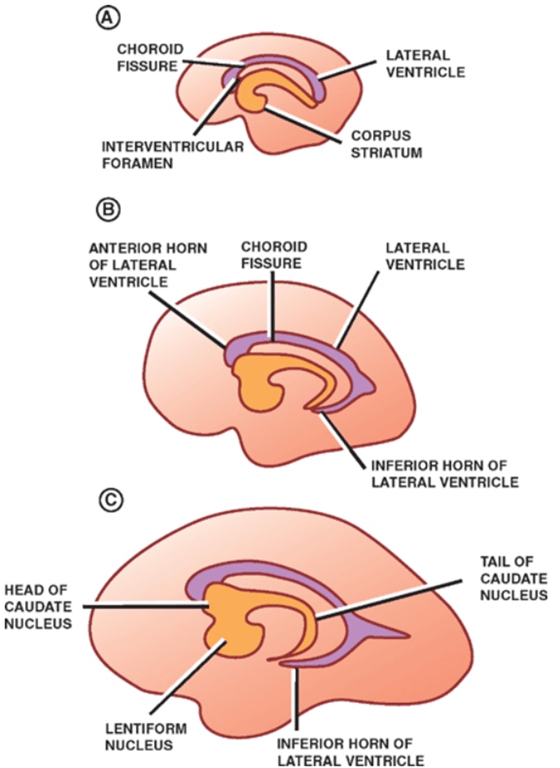
- The multiple directions through which the cerebral hemispheres expand account for its mature, C -shaped, configuration.
- Frontal lobe from anterior growth of the hemispheres
- Parietal lobe from lateral-superior growth
- Occipital and temporal lobes from posterior-inferior growth.
- Insula/insular cortex
- Slowly growing
- Is overgrown by the frontal, parietal, and temporal lobes and thus comes to lie deep in the lateral cerebral sulcus (sylvian fissure).
- C-shaped growth pattern found in
- Lateral ventricles
- Fornix
- Caudate nucleus
- Corpus striatum and internal capsule
- Corpus striatum
- Formed by
- Lentiform nucleus
- Putamen
- Globus pallidus
- Functionally and histologically distinct from the striatum
- Gives rise to the major efferents from the basal ganglia.
- Striatum
- Consist of (both are similar functionally and histologically)
- Putamen
- Caudate nucleus
- Receives all of the afferent input to the basal ganglia.
- Develop within the thick floor of the cerebral hemispheres, which undergo less lateral growth than the thin cortical walls. --> the striatum remains close to the midline of the brain, just lentiform nucleus lies ventrolateral to the caudate,
- Internal capsule
- Contains fibers headed to and from the cortex.
- Anterior limb of internal capsule separates the anterior limb of the internal capsule
- Posterior limb of the internal capsule separates the lentiform nucleus from the thalamus.
- Filled by the lateral ventricles
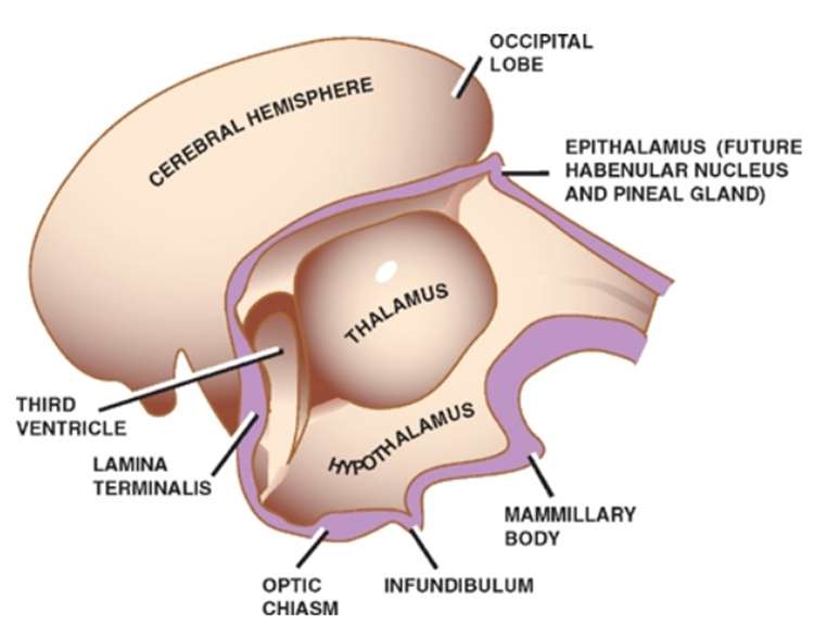
- Retinas and the pituitary gland are both derived from a combination of surface and neural ectoderm, the latter of which is derived from a downward evagination of the diencephalon.
- Develops into the
- Thalamus
- Hypothalamus
- Epithalamus
- optic cups
- Neurohypophysis
- With the associated central cavity forming the third ventricle
Telencephalon (rostrally)
Gives rise
1x median portion
2x lateral diverticula (telencephalic vesicles)
Anterior commissure
Hippocampal (commissure of fornix) commissure
Corpus callosum
Diencephalon (caudally)
Trapped mesenchyme between the developing cerebral hemispheres gives rise to the falx.
Mesencephalon (Midbrain)
- The mesencephalic flexure separates Mesencephalon from the rhombencephalon.
- The shrunken central cavity gives rise to the aqueduct.
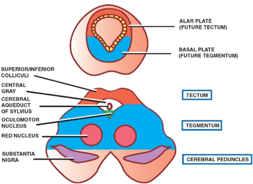
- Cells on the dorsal side of the neural tube (the alar plate) migrate dorsally, giving rise to the tectal plate, including the superior and inferior colliculi
- Others migrate ventrally to form the red nuclei and substantia nigra
- One of the least dramatic changes during development
- Central cavity of the midbrain: Cerebral aqueduct of Sylvius
- Neuroblasts from the alar plates migrate into
- Roof, or tectum , of the midbrain to form: The quadrigeminal plate
- inferior colliculi , which are concerned with audition,
- superior colliculi , which are concerned with visual reflexes.
- Central gray surrounding the aqueduct is also derived from neuroblasts of the alar plates.
- Neuroblasts from the basal plates
- give rise to several groups of neurons in the tegmentum of the midbrain:
- Oculomotor (III) and
- Trochlear (IV)
- Reticular nuclei,
- Red nuclei
- Substantia nigra .
- Two cerebral peduncles in the ventral midbrain contain cortical fibers descending to the brainstem and spinal cord.
Rhombencephalon (Hindbrain)
- Formed as the fourth ventricle expands, spreading its lateral walls open like the pages of a book.
- Local reabsorption of the roof of the 4th forms
- two lateral apertures (the foramina of Luschka)
- one median aperture (the foramen of Magendie)
- Pontine flexure divides hindbrain into
- The metencephalon develops into the pons and the cerebellum (with a contribution from the mesencephalon)
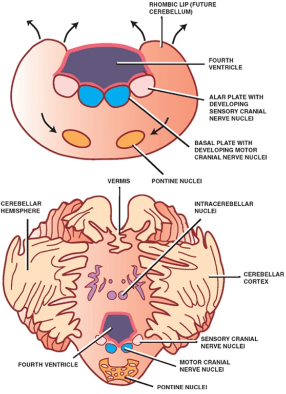
- Dorsal part develops into the cerebellum
- From the fusion of dorsolateral thickenings (rhomboid lips) of the metencephalon that overgrow the roof of the fourth ventricle.
- Rhomboid lips come together posteriorly in the midline to form the cerebellar vermis
- Peripherally migrating neuroblasts differentiate to form the cerebellar cortex
- Centrally migrating neuroblasts differentiate into the deep intracerebellar nuclei
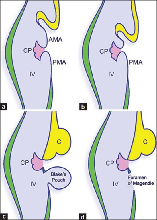
- Alar laminae along the lateral margins of the AMA become thickened to form two rhombic lips, which enlarge to approach each other and fuse in the midline dorsally (covering the rostral half of the 4th ventricle and overlapping the pons and the medulla) --> As the rhombic lips grow to form the cerebellar hemispheres and midline vermis, the AMA regresses by incorporation into the developing choroid plexus --> Growth and backward extension of the cerebellum pushes the choroid plexus inferiorly
- Folding, transverse fissure formation and foliation result in anterior lobe (cerebellar vermis and hemisphere above primary fissure), posterior lobe (vermis and hemispheres below primary fissure) and a flocculonodular lobe
- Development of the cerebellar cortex and deep nuclei (dentate, globose, emboliform, and fastigial) occurs as follows:
- Metencephalon consists of typical
- Ventricular layers
- The ventricular layer produces four types of neurons forming the mantle layer which will subsequently migrate to the cortex:
- Purkinje cells
- Golgi cells
- Basket cells
- Stellate cells
- Associated glia
- astrocytes including Bergmann glia, and oligodendrocytes
- Mantle layers
- Marginal layers
- Rhombic lips have started to form the cerebellum.
- Two additional layers form:
- an external germinal/granular layer derived from the rhombic lips, from which granular cells migrate inwards to form a new internal germinal layer between the ventricular and marginal layers (cells of the mantle layer have now dispersed into the marginal layer where they will form a distinct cortical pattern).
- External germinal layer also produces primitive nuclear neurons which also migrate inwards to form the deep cerebellar nuclei.
- Migration of granule cells takes place along Bergman (radial) glia.
- Purkinje cells migrate toward the cortex, it reels out an axon that maintains synaptic contact with neurons in the developing deep cerebellar nuclei.
- These axons will constitute the only efferents of the mature cerebellar cortex.
- From superficial to deep the cerebellum consists of:
- external granular layer (persists until approximately 15 months postnatally),
- Purkinje cell layer
- molecular layer (stellate, basket cells)
- granular layer (Golgi cells; granule cells and their parallel fibers),
- white matter (Mossy fibers from brainstem nulcei, climbing fibers from inferior olivary nucleus)
- deep cerebellar nuclei.
- The PMA expand like the finger of a glove forming blake's pouch.
- This is due to the 4th ventricle expanding caudally
- This Blake's pouch
- consists of ventricular ependyma surrounded by condensation of the mesenchymal tissues
- is initially a closed cavity that does not communicate with the surrounding subarachnoid space of the cisterna magna.
- The network between the vermis and the Blake's pouch progressively becomes condensed, whereas the other portions about the evagination become rarified resulting in permeabilization of the Blake's pouch to form the foramen of Magendie.
- The foramina of Luschka also probably appear late into the 4th month of gestation.
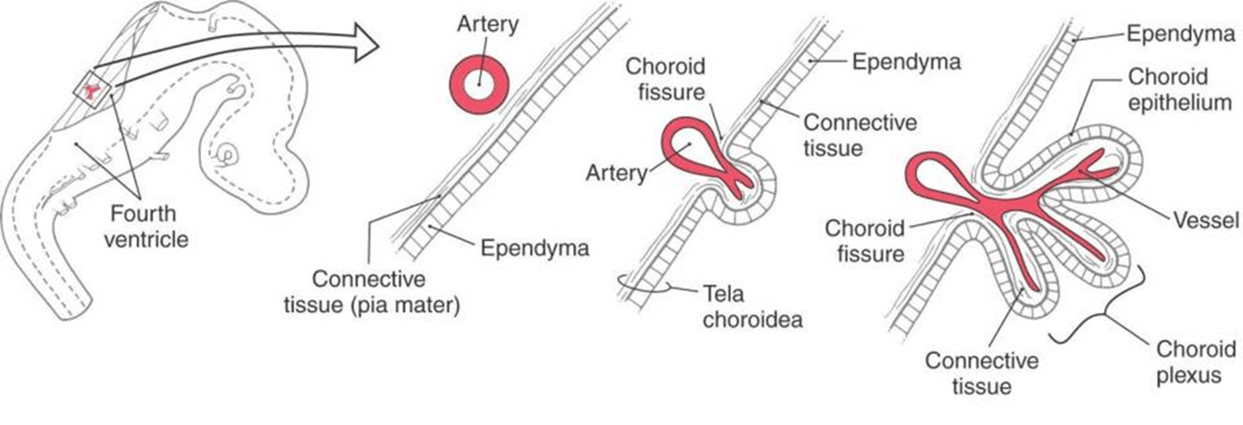
- The definitive tela choroidea of the 4th ventricle is form by: (From superior to inferior)
- Residual AMA
- Choroid plexus
- Residual PMA (i.e., residual rhombencephalic roof plate).
- Molecular layer
- Purkinje layer
- lamina dissecans (Disappears by 32 week)
- Internal granular layer
- Ventral part develops into the pons
- Development of the pons occurs ventral to the cerebellum in the ventral aspect of the metencephalon.
- Ventral pons
- Contains
- Pontine nuclei whose axons project to contralateral cerebellar cortices
- Dorsal pons
- Contains
- Cranial nerve nuclei
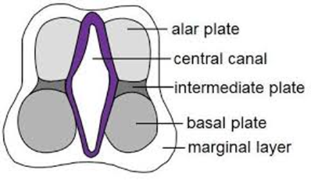
- Medially: Motor cranial nerve nuclei are derived from the basal plate
- Laterally: Sensory cranial nerve nuclei are derived from the alar plate.
- The myelencephalon develops into the medulla oblongata.
- Splaying of the rhombencephalon and stretching of the roof plate leads to the formation of the fourth ventricle
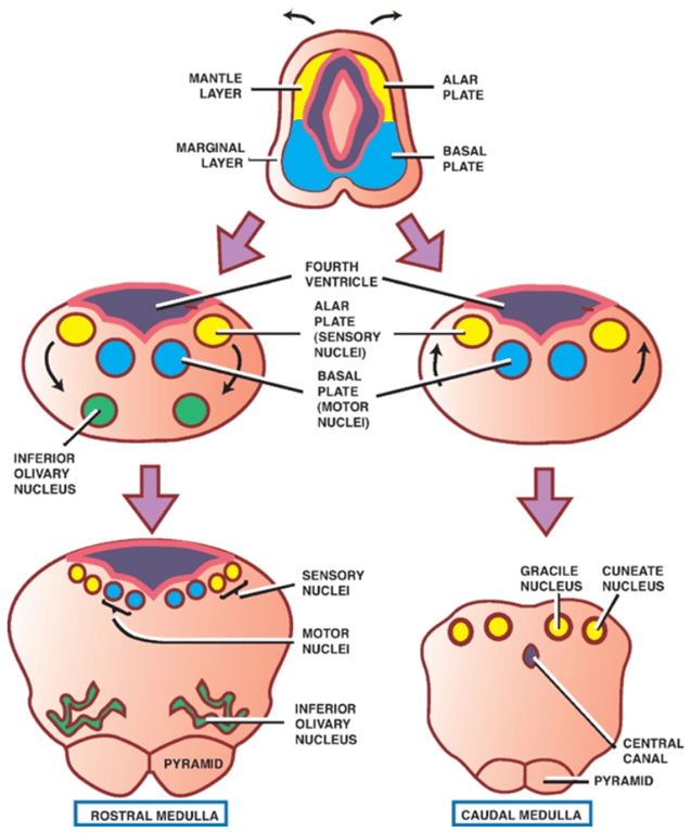
- Similarities between medulla vs spinal cord changes as the fourth ventricle expands resulting in
- Alar plates assume a lateral position in relation to the basal plates.
- Explains why sensory neurons (alar plates) lie lateral to motor neurons (basal plates) in the pons and medulla VS dorsal-ventral relation @ spinal cord
- Neuroblasts of the alar plate develop into sensory nuclei
- Some neuroblasts of the alar plate migrate ventrally to form isolated areas of gray matter:
- Inferior olivary nuclei
- Associated with the cerebellum,
- Gracile and cuneate nuclei
- Associated with the dorsal column-medial lemniscus tracts.
- Neuroblasts of the basal plate develop into motor nuclei.
- On the ventralmost aspect of the caudal medulla are the medullary pyramids, which contain the cortico-spinal tracts.
Metencephalon (the future pons and cerebellum)
cerebellum is formed
Bending of the pons (pontine flexure) → causes alar columns are splayed laterally → alar column eventually lie dorsolateral to the basal columns → this causes the roof plate of the developing 4th ventricle to be thin (The 4th ventricle is wide at its fold/waist and tapers superiorly and inferiorly (diamond shaped)) → Mesenchyme inserts itself into the roof fold and forms the plica choroidalis (choroid plexus precursor) (this divides the roof of the 4th ventricle into 2 cerebellar territory
Superiorly placed anterior membranous area (AMA)
Week 8
Week 12
Week 15
Inferiorly placed posterior membranous area (PMA))
Layers of the premature cerebellum
Myelencephalon (the future medulla)
 Made with Bullet