- The cranial sensory (afferent) and parasympathetic (visceral efferent) ganglia appear during the end of the 4th week and the beginning of the 5th week.
- Neurons in the cranial nerve sensory ganglia have a dual origin.
- Neural crest cells/Ectodermal placodes
- Nasal placodes
- The nasal placodes give rise to the primary neurosensory cells of the olfactory epithelium, and the axons of these cells form the olfactory nerve (I), which penetrates the olfactory bulb of the telencephalon.
- With some exceptions, the remaining cranial nerve sensory ganglia show a regular stratification with respect to their origin: the ganglia that lie closer to the brain (proximal) are derived from neural crest cells, whereas the neurons of ganglia lying farther from the brain (distal ganglia) are formed by placode-derived cells.
- Retinal placodes
- Otic placodes
- Epipharyngeal placodes
- The sensory ganglia associated with the second, third, fourth, and sixth pharyngeal arches are derived from the corresponding epipharyngeal placodes and from neural crest cells.
- Each of these nerves has both a
- Proximal sensory ganglion (e.g., superior ganglion of IX)
- Distal sensory ganglion (e.g., superior ganglion of IX).
- Pharyngeal arches
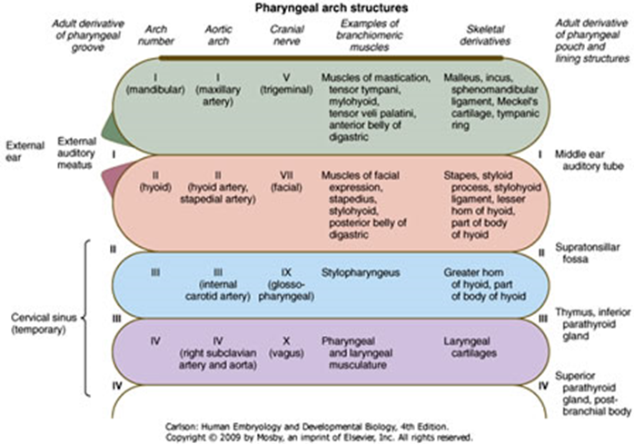
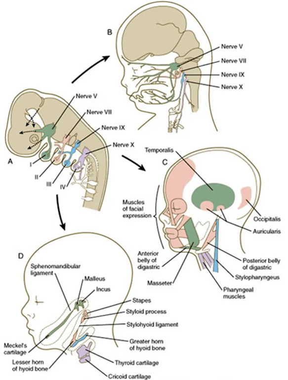
Arch | Nerve | Artery | Muscle | Bone |
1st (Mandibular) | V2 and V3 | Maxillary, ECA | Mastication Anterior belly of digastric Mylohyoid Tensor tympani Tensor veli palatini | Maxilla, mandible, zygoma, temporal bone, incus, malleus, Meckel's cartilage, sphenomandibular ligament |
2nd (Hyoid) | VII | Stapedial, hyoid | Muscles of facial expression Buccinator Platysma Stapedius Stylohyoid Posterior belly of digastric Auricular | Stapes, temporal styloid process, lesser horn and upper body of hyoid, stylohyoid ligament, Reichert's cartilage |
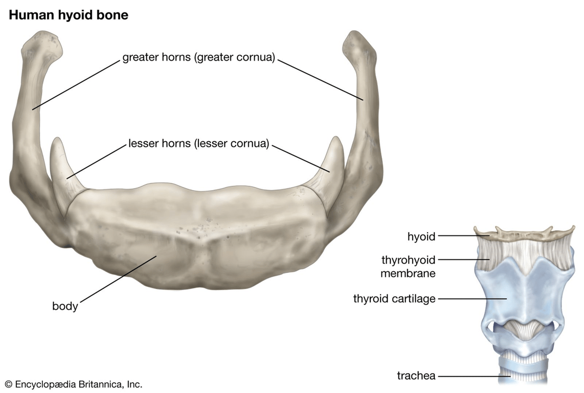
3rd | IX | CC/ICA | Stylopharyngeus | Greater horn and lesser body of hyoid, thymus, inferior parathyroids |
4th | X, superior laryngeal | Right: subclavian Left: aortic arch | Cricothyroid Intrinsic muscle of soft palate (except tensor veli palatine) | Thyroid cartilage, superior parathyroids, epiglottic cartilage |
6th | X, recurrent laryngeal | Right: pulmonary Left: pulmonary and ductus arteriosus | Intrinsic muscle of larynx (except cricothyroid) | Cricoid cartilage, arytenoid cartilage, corniculate cartilage, cuneiform cartilage |
 Made with Bullet