The presomitic stage
- Epiblastic cells from the embryonic plate caudal to the head process invaginate through the primitive streak to form mesoderm on each side of the neural plate
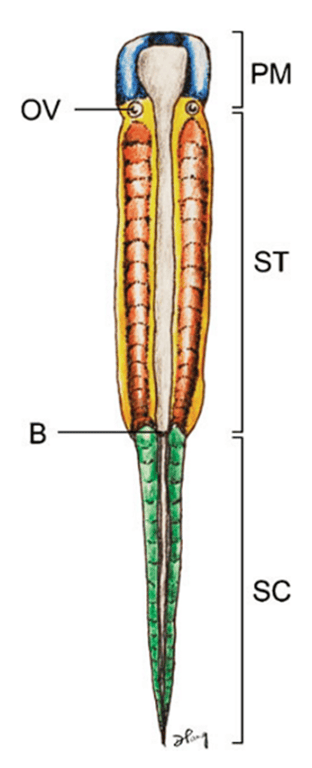
Vertebrate embryonic plate around gastrulation showing the three main regions of the body plan:
- Prechordal mesoderm (PM)
- Cranial to the otic vesicle (OV)
- forms most of the bones and muscles of the head and face without ever developing somites
- Somitic region of the trunk (ST)
- Between
- OV
- Blastopore (B)
- Forms the future anus
- Epiblast cells going through the primitive streak condense to form the parachordal mesoderm (aka presomitic mesoderm (PSM)/ segmental plate) which eventually forms the somites. Somites form the which will eventually give rise to the smooth muscle of the dermis, the axial musculature, the vertebral column, and support structures of the peripheral nervous system
- Caudal (tail) somitic region (SC)
- Caudal to the Blastopore
- After blastopore closure and complete regression of the primitive streak, the gastrulating region for the tail is restricted to a small cluster of cells called the tail bud, located at the caudal tip of the primitive streak.
- The tail bud functions as a blastema of undifferentiated cells
- The presomitic mesoderm and later somites of the caudal and tail regions are therefore not formed by epiblastic ingression but by progenitor condensation in situ.
Primary segmentation: somitogenesis
During somitogenesis, the loose mesenchymal cells of the PSM undergo transformation
into tightly apposed epithelial cells with definite polarity and orientation.
into tightly apposed epithelial cells with definite polarity and orientation.
- Somitogenesis begins soon after internalization of the prochordal (head) mesoderm and continues through subsequent production of the body axis
- first somite forms immediately caudal to the otic vesicle followed by sequential transformation such that a new pair of somites is regularly added in a rostrocaudal direction until a fixed species-specific number of somites is reached
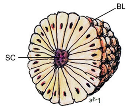
- A newly formed somite is a compact epithelial sphere (“somitomere”) composed of a single layer
of radially arranged cells with apices pointing towards a central lumen
(somitocoele, SC), which contains a few mesenchymal cells
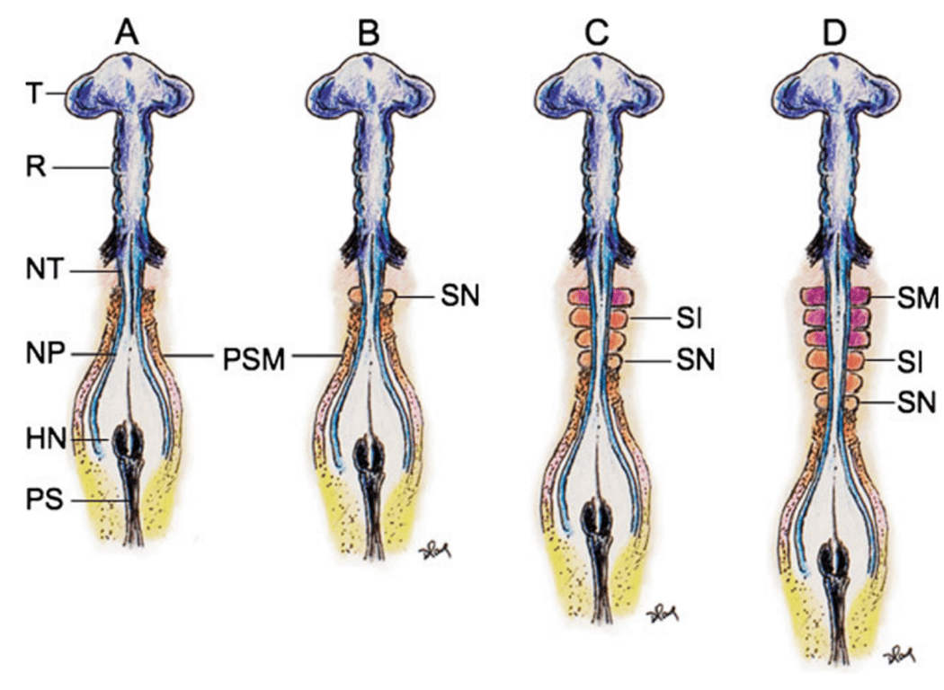
- (A) At very early gastrulation, the mostly unneurulated neural plate caudal to the prochordal
plate (head process) is flanked on each side by the presomitic (presegmented)
mesoderm, or PSM.
- (B) The first pair of somites are formed just caudal to the otic vesicles. The PSM column elongates
from addition of new cells from the caudal embryonic pole rostral to Hensen’s
node.
- (C) The new somites (SN) are formed in a rostrocaudal direction so that the older more matured
somites (SM) are rostral, i.e. closer to the cephalic end of the embryo.
- (D) The older matured somites (SM) have undergone dorsoventral differentiation (purple colour) into
sclerotome and dermomyotome. The intermediate-aged somites (SI) (orange colour)
are pre-differentiated units without dorsoventral specification. The PSM is always
at the most caudal end closest to Hensen’s node.
- Rostrocaudal sequential somitogenesis parallels progression of primary neurulation. HN
Hensen’s node, PS primitive streak, T telencephalon, R rhombencephalon, NT
neural tube, NP neural plate
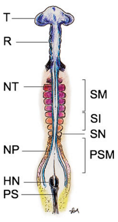
- The rostral most mesoderm consists of matured somites (SM) already having undergone dorsoventral differentiation into dermomyotome and sclerotome, flanking the formed neural tubes (NT); followed by the newer intermediate-aged predifferentiated epithelial somites (SI), the new somites (SN) and the presomitic mesoderm (PSM) on each side of the unneurulated neural plate (NP). HN Hensen’s node, PS primitive streak, T telencephalon, R rhombencephalon
- Caudal Cranial axial orientation is determined by the concentration of FGF8.
- Cranial somites has lower FGF8
- Caudal somites has higher FGF8
Differentiation of the somitic mesenchyme
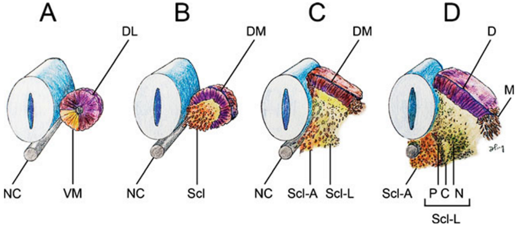
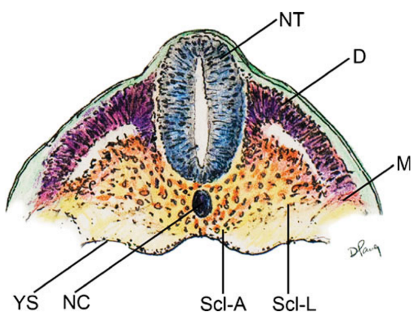
Within hours after its formation, each somite starts differentiating along its dorsoventral axis
- Cells from the ventromedial part (VM) --> Mesenchymal sclerotomes (Scl)
- lose their epithelial arrangement and migrate towards the notochord to form, with the luminal cells, the mesenchymal sclerotomes (Scl). Scl further divide into
- Axial scleretome (Scl-A) surrounding the notochord --> form vertebral column
- Lateral scleretome (Scl-L) flanking the perichordal axial sclerotome
- Scl-L forms a triangle next to the Scl-A. Each side of triangle forms
- pedicle (P),
- neural arch (N)
- the costal process (C)
- Cells from the dorsolateral part (DL) --> Dermomyotomes (DM)
- of the somite retain their epithelial arrangement to produce the dermomyotomes
- The dermomyotomes later subdivides into
- Dermatome (D)
- Dorsal placed
- immediately beneath the ectoderm
- Forms
- Dermis
- Smooth muscles
- Myotome
- Disaggregated cells between the dermatome and the sclerotomes remain closely packed as the myotome
- Form axial skeletal muscles
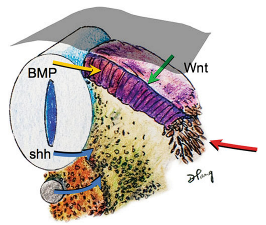
Ventral/dorsal axial orientation is determined by the concentration of FGF8.
- Expressions of sonic hedgehog gene (shh) (blue arrows) from the notochord and floor plate of the neural tube signal sclerotomal differentiation and ventral migration from the somite.
- Bone morphogenetic protein (BMP) (yellow arrow) expressed by the roof plate and Wnt signalling (green arrow) from the ectoderm (grey mantle) induce dermomyotome differentiation.
- Other lateralizing signals (red arrow) further encourage myotomal cell development
“Resegmentation” of the sclerotome
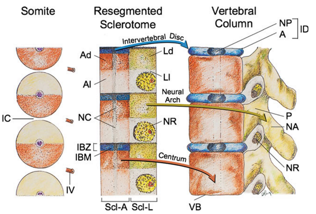
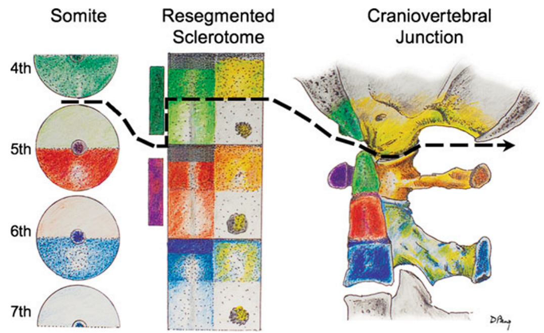
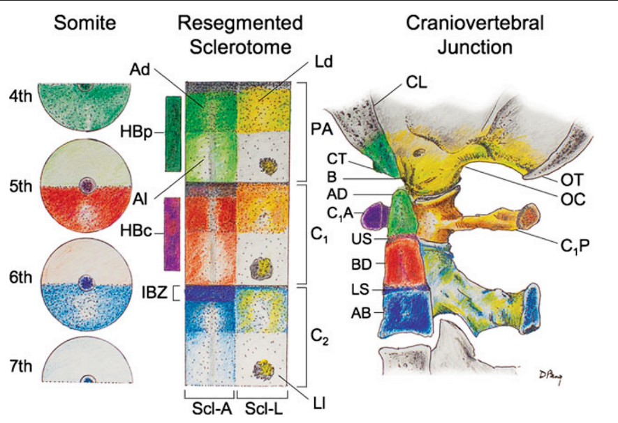
Resegmentation of somites to form sclerotomes and changes of sclerotomal primordia to mature
vertebral parts.
vertebral parts.
- During resegmentation, the sclerotome is formed from the caudal and rostral halves of two adjacent somites, such that the middle of the resegmented sclerotome lines up with the intersomitic cleft (IC).
- Both the axial sclerotome (Scl-A) and lateral sclerotome (Scl-L) develop dense and loose zones.
- The dense zone of the lateral sclerotome (Ld)
- becomes the
- neural arch (NA)
- pedicle (P)
- is attached to the rostral part of the vertebral body (VB) formed from chondrification of the loose (Al) and part of the dense zones (Ad) of the axial sclerotome.
- The rostral layer of the dense zone of the axial sclerotome soon forms the intervertebral boundary zone (IBZ) containing intervertebral boundary mesenchyme (IBM), which ultimately forms the annulus (A) and, together with notochord remnants (NC), the nucleus pulposus (NP) of the intervertebral disc (ID).
- The loose zone of the lateral sclerotome (Ll) does not form bone but promotes emergence of the nerve roots (NR).
- Thus, the neural arch is derived from a single somite but the vertebral body receives contributions from two adjacent somites. IV intersomitic vessel. Arrows indicate developmental fates of the sclerotomes
The severance line, which results in final cellular separation of the skull from the cervical spine, runs through the original resegmentation fronts of the adjacent somites 4 and 5, corresponding to the junction between the basion and apical segment of the dens in the axial proatlas, and between the exoccipital, or future occipital condyle, and the lateral mass of C1, derived from the lateral portion of the C1 resegmented sclerotome
 Made with Bullet