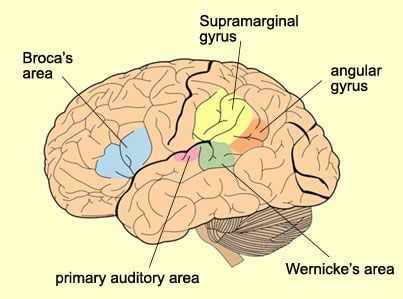
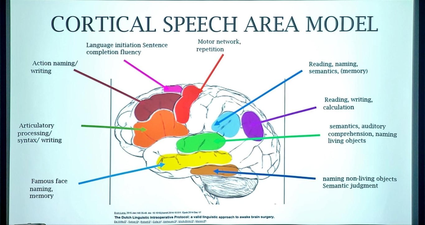
Areas involved with speech
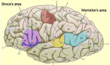
Broca's area
- Location
- Inferior frontal gyrus of dominant hemisphere
- Brodmann area 44
- Brodmann area 45
- Blood supply
- MCA
- Function
- Word formation (motor)
- Area 44
- Posterior part of the inferior frontal gyrus
- Seems to be involved in phonological processing and in language production as such;
- This role would be facilitated by its position close to the motor centres for the mouth and the tongue.
- Area 45
- (The anterior part of the inferior frontal gyrus)
- Seems more involved in the semantic aspects of language.
- Though not directly involved in accessing meaning
- Broca’s area therefore plays a role in verbal memory
- Selecting and manipulating semantic elements
- Deficit
- Expressive or motor aphasia
- Speech may be laboured and consist primarily of nouns, verbs or important adjectives. Speech takes on a telegraphic character.
- Difficulty with repetition
- Impairment in writing
Wernicke's area
- Location
- The posterior segment of the superior temporal gyrus in the dominant hemisphere.
- Broadmann's area 22
- Blood supply
- MCA
- Function
- Language comprehension: representation of phonetic sequences, regardless of whether the individual hears them, generates them himself or herself, or recalls them from memory.
- Deficit
- Fluent, grammatically correct speech with little meaning
- Poor comprehension
- Paraphasic errors:
- Calling a spoon a “fork” (semantic)
- Calling a spoon a “spood” (literal)
- Neologisms (or nonsense words)
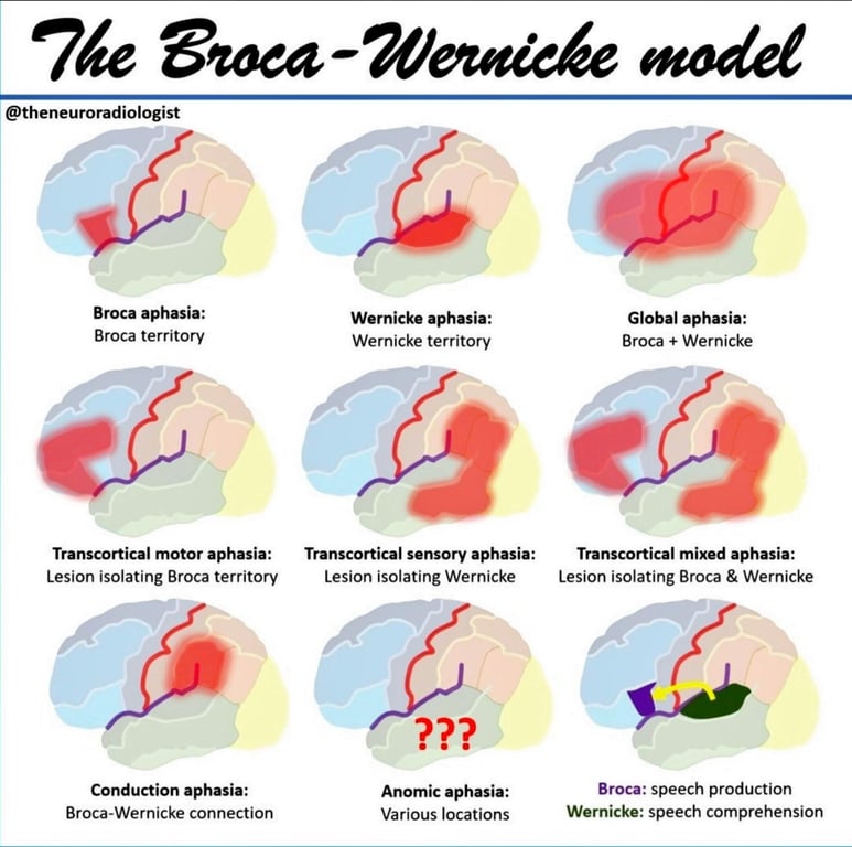
- Global Aphasia
- Both Wernicke’s and Broca’s areas damaged
- All aspects of speech and language are affected.
- Patients can say a few words at most and understand only a few words and phrases.
- Cannot carry out commands
- Cannot name objects
- Cannot read or write
- Cannot repeat words
Inferior parietal lobule: made up of
- Angular gyrus (area 39)
- Location
- Caudally located
- Which itself is bounded by the visual occipital areas (areas 17, 18, and 19),
- Function
- (Together with the posterior cingulate gyrus) seems more involved in semantic processing (process of finding meaning to words)
- Deficit
- Logopenic Primary Progressive Aphasia (lvPPA)
- Slowed speech with normal articulation,
- Impaired comprehension of sentence syntax
- Impaired naming of things
- Dyslexia (inability to read) without a deficit in understanding spoken language
- Supramarginal gyrus (area 40)
- Location
- Dorsally located
- Which arches over the end of the lateral sulcus, adjacent to the inferior portion of the somatosensory cortex
- Function
- Seems to be involved in phonological and articulatory processing of words
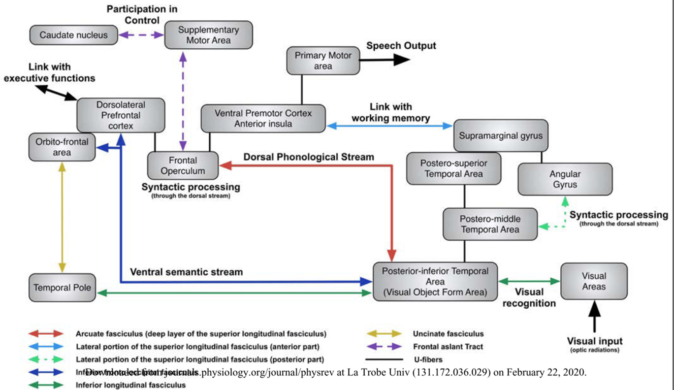
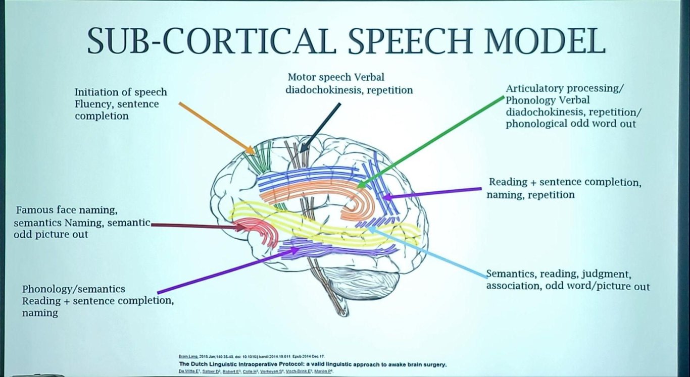
Connecting the different speech areas
- New model
- The dual-route model
- Ventral stream
- Which involves structures in the superior and middle portions of the temporal lobe, is involved in processing speech signals for comprehension.
- Bilaterally organised (although with important computational differences between the two hemispheres).
- This is in contrast to the typical view that speech processing is mainly left hemisphere dependent
- Dorsal stream
- Which involves structures in the posterior planum temporale region and posterior frontal lobe, is involved in translating acoustic speech signals into articulatory representations, which are essential for speech production.
- Strongly left-dominant.
- Arcuate fasciculus (aka: arcuate part of Superior longitudinal fasciculus)
- Old model
Wernicke-Geschwind model of language (1965)
graph TD
    A["Spoken word"] --> B["Area 41"]
    B --> C["Wernicke's area (contains<br>sound images of words)"]
    C --> D["Hear and comprehend word"]
graph TD
    A["Cognition"] --> B["Wernicke's area"]
    B --> C["Broca's area (stores motor<br>programs for speaking<br>words)"]
    C --> D["Facial area of motor cortex"]
    D --> E["Cranial nerves"]
    E --> F["Speak"]
graph TD
    A["Written word"] --> B["Area 17"]
    B --> C["Area 18, 19"]
    C --> D["Area 39 (angular gyrus)"]
    D --> E["Wernicke's area"]
    E --> F["Read"]
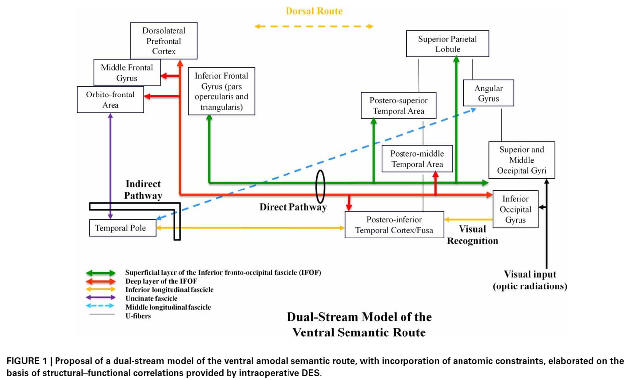
Language and semantics
- Has two streams that run in parallel no series
- Function:
- Processing visual information into meaning (semantics)
- Mapping sound to meaning: Auditory comprehension
- Local syntactic structure building
- Has a more bilateral distribution
- Formed by the
- IFOF
- Connects
- Occipital lobe + Superior parietal lobule + Fusiform gyrus (occipitotemporal gyrus) (FUSA) → frontal regions, (Inferior frontal gyrus + dorsolateral prefrontal cortex)
- Function:
- Verbal and non-verbal semantic processing
- Damage
- Left hemisphere: semantic paraphasias
- Left and right hemisphere: non-verbal semantic impairments
- Short form pyramid and palm tree test:
- Match a picture of a reference object (e.g., GLASSES) to the more associated of two objects depicted below it (e.g., target: EYE or distractor: EAR).
- Anterior ILF + Uncinate Fasciculus
- Connects:
- Anterior ILF: visual object form area (Right fusiform gyrus, FUSA) → the temporal pole (A semantic node enabling plurimodal integration of the multiple semantic-related signals originating from the unimodal systems) → to UF
- UF: temporal pole → the pars orbitalis of the inferior frontal gyrus
- Function:
- Semantic processing
- Proper name retrieval
- UF
- Lexical access
- Anterior ILF
- Function:
- Mapping visual information to articulation via visuophonological conversion
- Mapping sound to articulation (auditory to motor mapping)
- Syntactic processing
- Typically left-lateralised in right-handed individuals,
- Formed by the SLF which is formed by two subcomponents with distinct roles
- Secondary dorsal pathway:
- Formed by the classic Arcuate Fasciculus
- Aka: fronto-temporal part of the AF
- Connections
- The posterior temporal structures (mainly the middle and posterior fusiform/inferior temporal gyri) TO
- The inferior frontal gyrus (mostly the pars opercularis and triangularis (BA 44) – but also in the most ventral part of the posterior dorsolateral prefrontal cortex.
- Injury
- Conduction aphasia:
- Repetition disturbances along with phonemic paraphasia and its microstructural properties or its leftward lateralization predicts phonological abilities in healthy individuals.
- Formed by:
- Lateral part of the SLF III (called also horizontal component of the AF)
- IFC: inferior frontal cortex
- Connection
- Supramarginal gyrus + posterior part of the superior temporal gyrus (both of them receive feedback data from somatosensory and auditory regions) → ventral premotor cortex
- Function:
- Articulatory processing.
- Through the left occipital temporal pathway
- Pathway
- Occipital cortex (visual cortex) → posterior portion of ILF on the left hemisphere → fusiform area (gyrus) (visual word form area)
- Damage to
- Lower portion of the posterior ILF: Pure alexia
- Upper portion ILF: Anomia
Ventral subpathway:
Direct WM subpathway
Reference item | Target | Distractor |
Ink | Pen | Pencil |
Baby | Crib | Bed |
Drill | Screw | Nail |
Dog house | Dog | Cat |
Cheese | Mouse | Rabbit |
Tent | Camp fire | Radiator |
Web | Spider | Bee |
Matches | Candle | Light bulb |
Tree orchard | Apple | Onion |
Mice | Cat | Dog |
Pillow | Bed | Chair |
Glasses | Eye | Ear |
Wood | Saw | Hammer |
Curtain | Window | Door |
Indirect subpathway
Dorsal subpathway:
Deep component
graph LR
     A[Temporal cortex] -- Arcuate fasciculus --> BA44
Superficial component
graph LR
    A[Temporal cortex] -- IFC--> B[Premotor cortex]
		A -- SLF -->B
Naming pictures
graph TD
A["Concept (your concept of a<br> dog)"] --> B["Lemma retrieval<br> (abstract word form containing<br> information about meaning)<br> stored in the left middle<br> temporal gyrus, retrieved on<br> the basis of meaning- concept<br> activates lemmas and co-<br> activates related lemmas<br> (cat/walk/bark)"]
B --> C["Lexeme retrieval (Stored in<br> the posterior part of the left<br> superior and middle temporal<br> gyrus)"]
C --> D["Phonological encoding (left<br> inferior frontal gyrus)"]
D --> E["Programme articulation<br> (Left inferior frontal gyrus)"]
E --> F["Articulation (in bilateral<br> motor and sensory cortices)"]

Assessing speech
Summary
---
title: Normal
---
graph TD
    A[Hearing]--> B["Wernicke's Area<br>('Recognising sounds'<br>Dominant hemisphere<br>supramarginal gyrus &<br>angular gyrus)"]
    B--> C["Concept Area<br>(Understanding words)"]
    B-- Arcuate fasciculus --> D["Broca's Area (Br 44:<br>Dominant hemisphere<br>between pars operculairs<br>and pars triangular)"]
    C--> D
    D--> E["Voice Production<br>and Articulation"]

    classDef crossStyle fill:#ffdddd,stroke:#ff0000,stroke-width:4px;
    class z crossStyle;
3 main areas
- Quality
- Dysarthria
- Neurologic damage to the motor components of speech, which may involve any or all of the speech processes, including respiration, phonation, articulation, resonance, and prosody
- Dysphonia
- Disordered sound production at the level of the larynx (hoarseness)
- Content
- Expressive function
- Broca's area
- Understanding:
- Receptive function
- Wernicke's area
---
config:
  layout: dagre
---
graph TD
    A[Types of Aphasia] --> B{Fluent?}
    B -->|No| C{Comprehends?}
    C -->|No| D{Repeats?}
    D -->|No| E["Global aphasia (Large territory infarct)"]
    D -->|Yes| F["Mixed transcortical aphasia (Watershed Infarct affecting speech areas)"]
    C -->|Yes| G{Repeats?}
    G -->|No| H["Broca's aphasia (Inferior frontal lobe. Telegraphic speech)"]
    G -->|Yes| I["Transcortical motor aphasia (Frontal white matter lesion)"]
    B -->|Yes| J{Comprehends?}
    J -->|No| K{Repeats?}
    K -->|No| L["Wernicke's aphasia (Posterior lesion. Paraphasias and neologisms)"]
    K -->|Yes| M["Transcortical sensory aphasia (White matter underlying Wernicke’s area)"]
    J -->|Yes| N{Repeats?}
    N -->|No| O["Conduction aphasia (Arcuate fasciculus damage)"]
    N -->|Yes| P["Anomic aphasia (Isolated word finding deficit; least localized)"]

    linkStyle 0 stroke:White,stroke-width:4px
    linkStyle 1 stroke:red,stroke-width:4px
    linkStyle 2 stroke:red,stroke-width:4px
    linkStyle 3 stroke:red,stroke-width:4px
    linkStyle 4 stroke:green,stroke-width:4px
    linkStyle 5 stroke:green,stroke-width:4px
    linkStyle 6 stroke:red,stroke-width:4px
    linkStyle 7 stroke:green,stroke-width:4px
    linkStyle 8 stroke:green,stroke-width:4px
    linkStyle 9 stroke:red,stroke-width:4px
    linkStyle 10 stroke:red,stroke-width:4px
    linkStyle 11 stroke:green,stroke-width:4px
    linkStyle 12 stroke:green,stroke-width:4px
    linkStyle 13 stroke:red,stroke-width:4px
    linkStyle 14 stroke:green,stroke-width:4px
Types of dysphasia
- Fluent (receptive): problems understanding
- Wernicke's ('fluent aphasia'/posterior aphasia)
- Impairment of the ability to understand the meaning of spoken words
- Abnormal but fluent speech
- Sentences do not hang together and irrelevant words intrude-sometimes to the point of jargon, in severe cases.
- Reading and writing are often severely impaired.
- Conduction
- Transcortical sensory
- Nominal
- Nonfluent (expressive): Trouble using words and sentences
- Broca's (non fluent form)
- Speech output is severely reduced and is limited mainly to short utterances of less than four words.
- Vocabulary access is limited and the formation of sounds by persons with Broca's aphasia is often laborious and clumsy.
- Understand speech relatively well and be able to read,
- Limited in writing.
- Global aphasia:
- Struggle with both using words and understanding
- Cannot read nor write.
- Most severe form of aphasia,
- Patients who can produce few recognizable words and understand little or no spoken language.
- Transcortical motor
Aphasia
- Define: Term used to describe an acquired loss of language that causes problems with any or all of the following:
- Speaking
- Listening
- Reading
- Writing
- Reading and writing are more impaired than talking or understanding.
- Anomic aphasia
- This term is applied to persons who are left with a persistent inability to supply the words for the very things they want to talk about-particularly the significant nouns and verbs.
- As a result their speech, while fluent in grammatical form and output is full of vague circumlocutions and expressions of frustration.
- Understand speech well
- Read well
- Difficulty writing
- Primary Progressive Aphasia (PPA)
- Pathology
- Normal role for the site of initial degeneration → Eventually problems spread throughout the broader language network
- Language capabilities become slowly and progressively impaired.
- Subtypes include
- Nonfluent primary progressive aphasia (nfvPPA),
- Semantic variant primary progressive aphasia (svPPA)
- Logopenic primary progressive aphasia (lvPPA).
- Unlike other forms of aphasia that result from stroke or brain injury, PPA is caused by neurodegenerative diseases, such as
- Alzheimer's Disease
- Frontotemporal Lobar Degeneration.
- PPA results from deterioration of brain tissue important for speech and language.
- Although the first symptoms are problems with speech and language, other problems associated with the underlying disease, such as memory loss, often occur later.
- Mixed non-fluent aphasia
- Sparse and effortful speech, resembling severe Broca's aphasia.
- However, unlike persons with Broca's aphasia, they remain limited in their comprehension of speech and do not read or write beyond an elementary level.
Examination
- Inspection
- Look for any scars, tracheostomy, PEG
- Ask if the patient is right or left handed
- Aphasia:
- What is your name and address? [tests understanding and fluency]
- Give command: with your right hand, touch your nose [tests understanding]
- Assess whether speech is spontaneous
- Assess word finding:
- Name all the animals you can think of in 1 minute
- Name these 3 objects
- Assess repetition:
- Repeat: "the sun is shining"
- If able to repeat then wernicke’s area, broca’s area and arcuate fasciculus is not injured
---
title: Transcortical motor aphasia (Normal comprehension, Non-fluent speech, Normal repetition)
---
graph LR
    A[Hearing]--> B["Wernicke's Area<br>('Recognising sounds'<br>Dominant hemisphere<br>supramarginal gyrus &<br>angular gyrus)"]
    B--> C["Concept Area<br>(Understanding words)"]
    B-- "<span style="color: white;">Arcuate fasciculus</span>" --> D["Broca's Area<br>(Br 44: Dominant<br>hemisphere between<br>pars operculairs and<br>pars triangularis)"]
    C--> D
    D--> E["Voice Production<br>and Articulation"]

    classDef crossStyle fill:#ffdddd,stroke:#ff0000,stroke-width:4px;
    class Z crossStyle;
	  linkStyle 3 stroke:#ff0000,stroke-width:4px,color:#ff0000
---
title: Transcortical sensory aphasia (Poor comprehension, fluent but meaningless speech, Normal repetition)
---
graph LR
    A[Hearing]--> B["Wernicke's Area<br>('Recognising sounds' Dominant hemisphere<br>supramarginal gyrus &<br>angular gyrus)"]
    B--> C["Concept Area<br>(Understanding words)"]
    B-- "<span style="color: white;">Arcuate fasciculus</span>" --> D["Broca's Area (Br 44:<br>Dominant hemisphere<br>between pars operculairs<br>and pars triangularis)"]
    C--> D
    D--> E["Voice Production<br>and Articulation"]

    classDef crossStyle fill:#ffdddd,stroke:#ff0000,stroke-width:4px;
    class Z crossStyle;
	  linkStyle 1 stroke:#ff0000,stroke-width:4px,color:#ff0000
---
title: Conductive aphasia (Normal comprehension, no repetition)
---
graph LR
    A[Hearing]--> B["Wernicke's Area<br>('Recognising sounds'<br>Dominant hemisphere<br>supramarginal gyrus &<br>angular gyrus)"]
    B--> C["Concept Area<br>(Understanding words)"]
    B-- "<span style="color: red;">Arcuate fasciculus</span>" --> D["Broca's Area<br>(Br 44: Dominant<br>hemisphere between pars<br>operculairs and<br>pars triangularis)"]
    C--> D
    D--> E["Voice Production<br>and Articulation"]

    classDef crossStyle fill:#ffdddd,stroke:#ff0000,stroke-width:4px;
    class Z crossStyle;
	  linkStyle 2 stroke:#ff0000,stroke-width:4px,color:#ff0000
- Types of Aphasia:
- Global aphasia = lesion in both Broca's and Wernicke's areas
- Nominal aphasia = lesion in the angular gyrus
- Expressive dysphasia = lesion in the inferior frontal gyrus
- Receptive dysphasia = lesion in the supramarginal gyrus of the temporal lobe
---
title: Broca's aphasia (Normal comprehension, Non-fluent speech, no repetition)
---
graph LR
    A[Hearing]--> B["Wernicke's Area<br>('Recognising sounds'<br>Dominant hemisphere<br>supramarginal gyrus &<br>angular gyrus)"]
    B--> C["Concept Area<br>(Understanding words)"]
    B-- "Arcuate fasciculus" --> D["Broca's Area (Br 44:<br>Dominant hemisphere<br>between pars operculairs<br>and pars triangular)"]
    C--> D
    D--> E["Voice Production and<br>Articulation"]

    classDef crossStyle fill:#ffdddd,stroke:#ff0000,stroke-width:4px,color:#ff0000;
    class D crossStyle;
---
title: Wernicke's aphasia (Poor comprehension, fluent but meaningless speech, no repetition)
---
graph LR
    A[Hearing]--> B["Wernicke's Area ('Recognising<br>sounds' Dominant hemisphere<br>supramarginal gyrus &<br>angular gyrus)"]
    B--> C["Concept Area<br>(Understanding words)"]
    B-- Arcuate fasciculus --> D["Broca's Area (Br 44:<br>Dominant hemisphere<br>between pars operculairs<br>and pars triangular)"]
    C--> D
    D--> E["Voice Production<br>and Articulation"]

    classDef crossStyle fill:#ffdddd,stroke:#ff0000,stroke-width:4px,color:#ff0000;
    class B crossStyle;
- Dysphonia:
- Ask the patient to cough
- Say 'eeeeeee'
- Dysarthria (spastic/extrapyramidal/cerebellar/lower CN):
- Say "Yellow lorry"
- Say "Baby hippopotamus"
- Examine other functions in the frontal and parietal lobe
- Check for field deficit
- Check for motor weakness
- Cranial nerve exam
- Examine reading and writing
Clinical
Supplementary Motor Area Language Syndrome
- Anterior aspect of the left superior frontal gyrus:
- Suggested that the SMA has various superordinate control functions during speech communication and language reception, especially with increased task demands.
- SMA is subdivided into:
- Posterior region: Predominantly motor-related functions (SMA proper)
- Anterior part (pre-SMA): Involved in higher-order cognitive control mechanisms
- Intra-op symptoms:
- More difficult to start talking or appearing mute
- Single word responses rather than sentences
- Use of simple words rather than more complex or specific ones
- Talking less
- Less animated (lacking facial expression and gesture)
- Does not typically affect comprehension of language
- Post-op:
- "They don’t seem like they want to talk to me, they’re not talking much and don’t smile when they see me"
- Recovery is usually complete; improvement in 1st week but 2-9 weeks for complete recovery
 Made with Bullet