Surface anatomy of occipital squama
Trapezoidal-shaped bone is externally convex and internally concave.
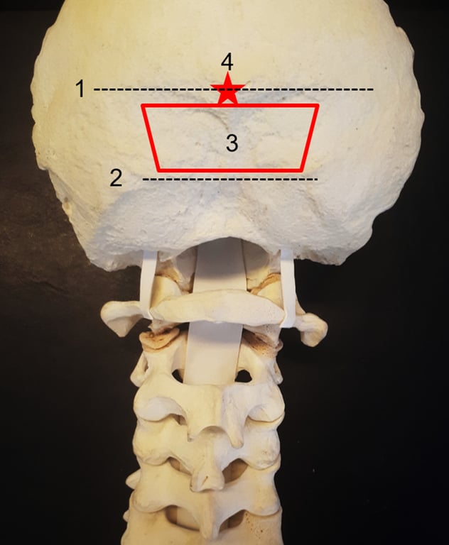
- Superior nuchal line—insertion of the trapezius, splenius capitis, and sternocleidomastoid muscle.
- The transverse sinus and the torcula are internal to this line.
- Inferior nuchal line—insertion of the obliquus capitis superior, rectus capitis posterior major, and rectus capitis posterior minor muscle. We generally perform, when necessary, decompression below this area.
- The “red trapezium” corresponds to the area of preference to insert the occipital screws
- just below the superior nuchal line to avoid a prominent plate, in the midline, where the occipital squama is thicker.
- The “red star” is the external occipital protuberance (EOP), which corresponds to the confluence of the dural sinuses (torcula). inion is the most prominent projection of the EOP
- EOP thickness ranged from 7.4 to 22.3 mm (mean of 14 mm); 1 cm below the EOP, the thickness ranged from 2.9 to 13.4 mm. → must look at scan and measure due to the great variable thickness between patients
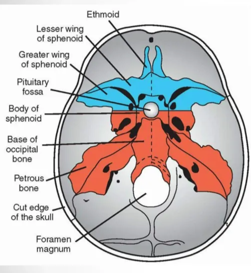
4 parts of occipital bone
- Each part has two surfaces: upper or external and lower or internal.
Basilar Part
- This quadrilateral part of the bone is adjacent to the petrous part of the temporal bone and anterior to the foramen magnum. It has an upper surface and a lower surface.
- During adolescence, the upper surface of the basilar part articulates with the sphenoid bone to form the clivus. The lower surface features the pharyngeal tubercle, where the superior pharyngeal constrictor muscle and fibrous pharyngeal raphe insert. The other muscles attached to the lower surface are rectus capitis anterior and longus capitis.
- The clivus is that part of the skull base situated between the foramen magnum and the dorsum sellae.
- Formed from sphenoid and occipital bones.
- The petrooccipital fissure forms the anterior lateral margin of the clivus, while the synchondrosis between the basioccipital and exoccipital bones forms the posterior lateral margins.
- Normal fat signals in adult (late teens) in MRI.
Squamous Part
- It is the largest of all four parts and contains internal and external surfaces. This part features the external occipital protuberance, a bony prominence in the middle of the outer surface.
- The trapezius muscle attaches here. The external surface also has three curved lines, called the nuchal lines. They are:
- Supreme nuchal line: The epicranius muscle and epicranial aponeurosis originate from here.
- Superior nuchal line: Running inferior to the squamous part, it serves as a site of origin for the trapezius, splenius capitis, and sternocleidomastoid muscles.
- Inferior nuchal line: Found further inferior to the superior nuchal line, this is where the semispinalis capitis muscle gets inserted.
Condylar Parts (2 parts)
- The condylar parts are commonly known as the lateral parts of the occipital bone, as they are found lateral to the foramen magnum. Each of the two condylar parts contains an upper and lower surface. It features two kidney-shaped prominences called occipital condyles that form articulation with the first cervical vertebra (C1), thus giving rise to the atlanto-occipital joint.
- The condylar canals are located just behind the condyles, through which the condylar emissary veins pass. The canals also connect the external vertebral venous plexuses with the sigmoid sinuses. The hypoglossal canal lies on the inferior surface of the condylar part through which the hypoglossal nerve leaves the cranium.
Margin of the occiput
- Anterior Margin
- Posterior surface of the clivus
- Lateral Boundaries
- Superior: Posterior surface of the petrous part of the temporal bone
- Inferior: Condylar part of the occipital bone
- Posterior Margin
- Mastoid part of the temporal bone
- Squamous part of the occipital bone
- Central Structure
- Foramen magnum
Occipital bone embryonal origin
- The occipital bone has a double origin:
- Endochondral for its basilar part surrounding the foramen magnum
- Membranous for its squamous (flat) part.
 Made with Bullet