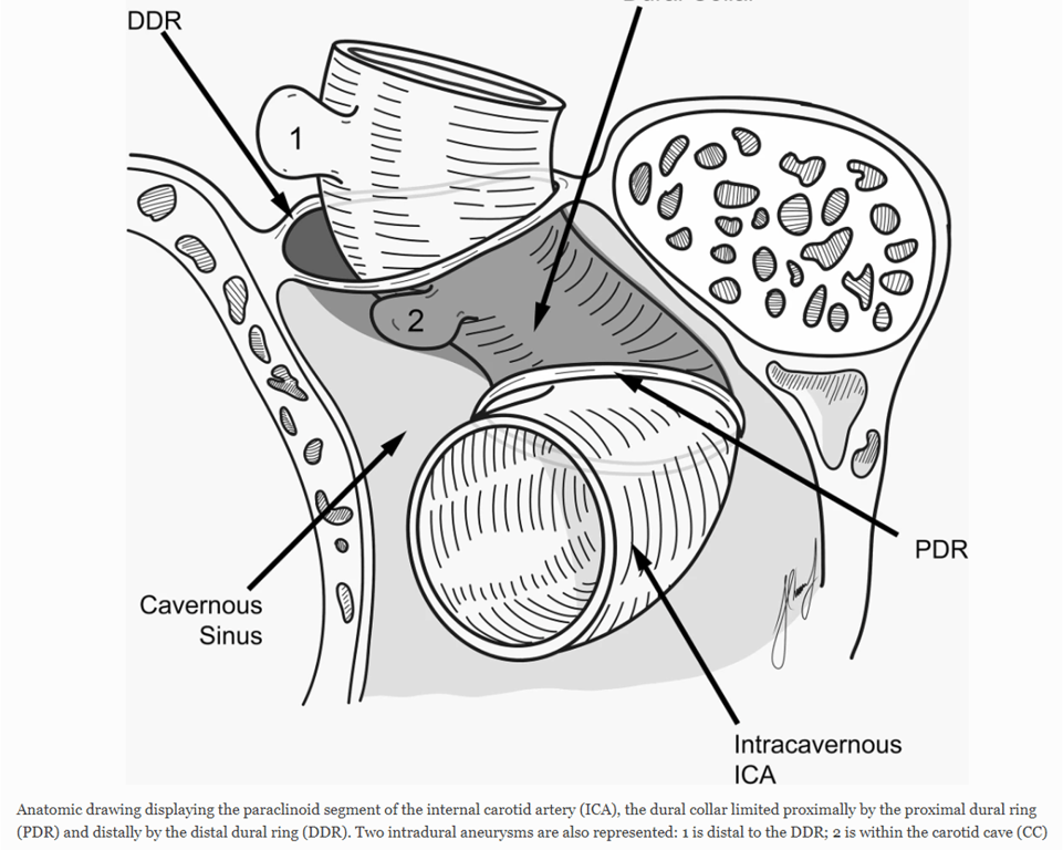
- The clinoidal carotid segment is a transitional segment between the cavernous sinus before the ICA exits through the distal ring and enters the subarachnoid space
- Two rings are fused posteriorly, they are variably separated anteriorly, giving rise to two regions medially and laterally.
- The lateral region may be considered to be extradural and extracavernous.
- On the medial side, the space between a redundant portion of distal dural ring and ICA is termed the carotid cave.
- Although usually extradural, rupture of carotid cave aneurysm extending superiorly out of the cave may result in subarachnoid haemorrhage
- Distal dural ring sits inclined posterior-medially and is continuous medially with the falciform ligament and with the dura overlying the superior-medial aspect of the anterior clinoid process
- Surgically understanding of this region is important as access to proximal aneurysms at or near the origin of the ophthalmic artery will require an anterior clinoidectomy
- Distal ring is not visible radiologically and its location can only be estimated.
- Clinically this region is important as the distal dural ring differentiates aneurysms that cause subarachnoid haemorrhage versus those that cause carotico-cavernous fistulae.
- Anatomical Exposure and Regions
- The optic strut is exposed in the anterior part of the clinoidal triangle.
- The clinoid segment is exposed in the midportion.
- The roof of the cavernous sinus is exposed in the posterior part.
- The posterior bend of the internal carotid artery (ICA) and the origin of the meningohypophyseal trunk are exposed in the infratrochlear triangle.
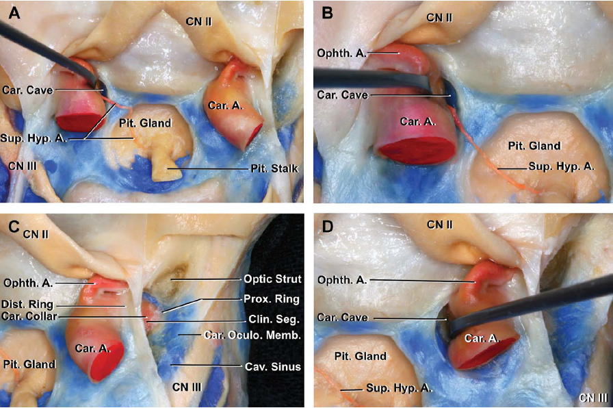
- Dural Rings and Membranes
- The upper margin of the clinoid segment is surrounded by the upper dural ring.
- The upper dural ring is formed by the dura extending medially from the upper surface of the anterior clinoid.
- The lower dural ring defines the lower margin of the clinoid segment.
- The dura on the lower margin of the anterior clinoid is referred to as the carotidoculomotor membrane.
- The carotidoculomotor membrane separates the lower surface of the anterior clinoid from the upper surface of the oculomotor nerve.
- This membrane extends medially to form the lower dural ring.
- Internal Carotid Artery (ICA) and Branches
- The meningohypophyseal trunk arises near the posterior bend of the ICA.
- The meningohypophyseal trunk gives rise to the tentorial and dorsal meningeal arteries.
- The inferolateral trunk arises from the horizontal segment of the intracavernous carotid.
- Cranial Nerve Pathways
- The abducens nerve passes through Dorello’s canal.
- The abducens nerve passes between the lateral surface of the intracavernous carotid and the medial side of the ophthalmic nerve.
- The inferolateral trunk passes above the abducens nerve.
- The right middle clinoid base is located at the center of the C-shaped anterior bend of the cavernous carotid artery.
- The middle clinoid is covered by the periosteal layer of the dura mater (yellow arrow).
- It protrudes into the cavernous sinus, with its base just below the projection level of the clinoid segment of the internal carotid artery according to intracranial dissections.
- The proximal dural ring is not identifiable from an endonasal view on the medial side of the artery.
- The caroticoclinoid ring is an osseous bridge formed by the fusion of the anterior clinoid process (ACP) and the middle clinoid process.
- The middle clinoid process itself is a prominence located on the medial side of the terminal part of the carotid sulcus, medial to the tip of the ACP.
- This fusion creates a bony ring or canal—the caroticoclinoidal foramen—through which the carotid segment of the ICA passes through
- The anterior clinoid process has been surgically removed.
- The III, IV, and V cranial nerves have been resected, along with both the anterior and posterior bends of the cavernous carotid artery.
- In the presence of a caroticoclinoid ring, the carotidoculomotor membrane continues posterior to the carotid artery below the osseous ring and continues the proximal dural ring (yellow dotted lines) all the way to the medial wall of the cavernous sinus.
- The osseous ring forms part of the roof of the cavernous sinus.
- The distal dural ring is represented by blue dotted lines.
- The left anterior clinoid has been removed.
- The carotidoculomotor membrane forms the proximal dural ring when it meets the carotid medially.
- Blue silicon, representing the venous filling in the cavernous sinus, has been partially removed below the distal dural ring posteriorly.
- The proximal dural ring is not evident in the posterior and medial aspect of the carotid.
- In some specimens, there is a carotico clinoid ligament between the anterior clinoid and middle clinoid (when present) or medial border of the carotid sulcus at the level of the anterior bend of the carotid artery (when the middle clinoid is absent).
- Illustration of the DCS from the endoscopic endonasal view.
- Illustration of the DCS from the microsurgical posterior view; the ACP has been removed.
- Posterosuperior view of the sella.
- The dorsum and posterior clinoid have been removed to expose the posterior lobe of the pituitary, which was hidden below the dorsum.
- The abducens nerve is exposed below the petrosphenoidal ligament.
- The trigeminal nerve has been reflected forward to expose the petroligand ligament, which extends above the internal carotid artery just proximal to the artery’s entry into the cavernous sinus.
Stepwise microscopic dissection: superior to inferior
- Showing the intracranial correlation of the recesses and referenced points in the sellar wall of the sphenoid sinus, superior intracranial view.
- After brain removal, the dura mater covering the right anterior clinoid process has been removed.
- The 1-mm perforations are enhanced with green circles for clarity.
- The right anterior clinoid process has been drilled, view of the 3 reference points (6, 7, 8) of the lateral opticocarotid recess corresponding to the optic strut.
- Note the point corresponding to the deepest and middle part of the tuberculum recess (1).
- The proximal and distal dural rings and the carotidoculomotor membrane are exposed.
- The middle clinoid process is not visualized as it is located below the level of the posterior junction of the proximal and distal dural rings at the tip of the anterior clinoid.
- The dural rings have been incised to retract laterally the right carotid artery, and the periosteal layer of the dura mater has been removed.
- The medial opticocarotid point (4) and caroticosellar point (5) are exposed with the middle clinoid (yellow dotted line).
- The caroticosellar and medial opticocarotid points are superior to the middle clinoid.
- Bilateral dissection showing both junction points and bilateral prominence of the middle clinoid processes.
 Made with Bullet