Anatomy
- C1 uncovertebral (Lushka’s) joints
- are formed by uncinate processes that extend upward from the lateral margin of the superior surface of the vertebral body and limit lateral flexion/guide flexion-extension.
- C1 weakest where the anterior and posterior arches connect to the lateral masses
- The C1 lateral mass screw is partially threaded to reduce irritation of the C2 nerve root and subsequent neuropathic pain.
- The atlas consists of two thick lateral masses situated at the anteromedial part of the ring, which are connected in front by a short anterior arch and posteriorly by a longer curved posterior arch.
- The superior articular facet is an oval, concave facet that faces upward and medially to articulate with the occipital condyle.
- The inferior articular facet is a circular, flat, or slightly concave facet that faces downward, medially, and slightly backward and articulates with the superior articular facet of the axis.
- The medial aspect of each lateral mass has a small tubercle for the attachment of the transverse ligament of the atlas.
- The transverse process projects from the lateral masses.
- The upper surface of the posterior arch adjacent to the lateral masses has paired grooves in which the vertebral arteries course.
A, superior view;
- Inferior view
- Anterior view
- Posterior view
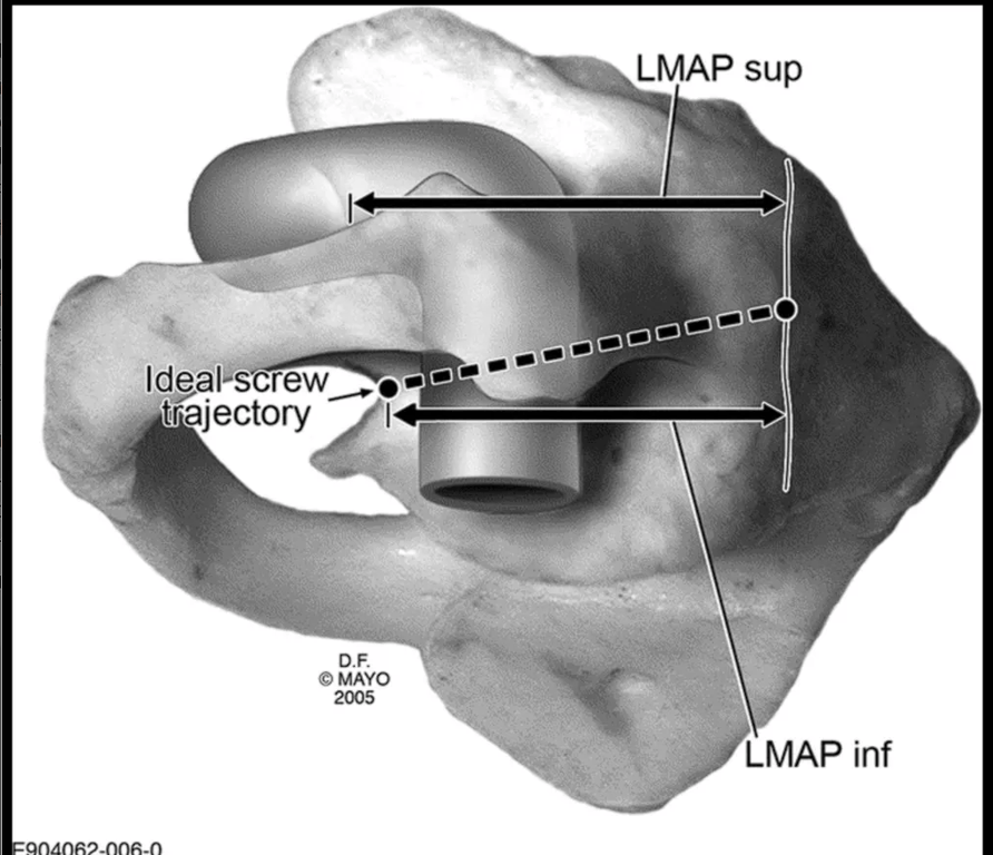
- Lateral view of C1 demonstrating the ideal orientation of screw placement within the sagittal plane (10°–15° caudad to cephalad).
- Note the starting position below the posterior ring confluence with the lateral mass.
- The arrows represent the following dimensions:
- LMAPsup, lateral mass anteroposterior dimension superior to the posterior ring insertion;
- LMAPinf, lateral mass anteroposterior dimension inferior to the posterior ring insertion.
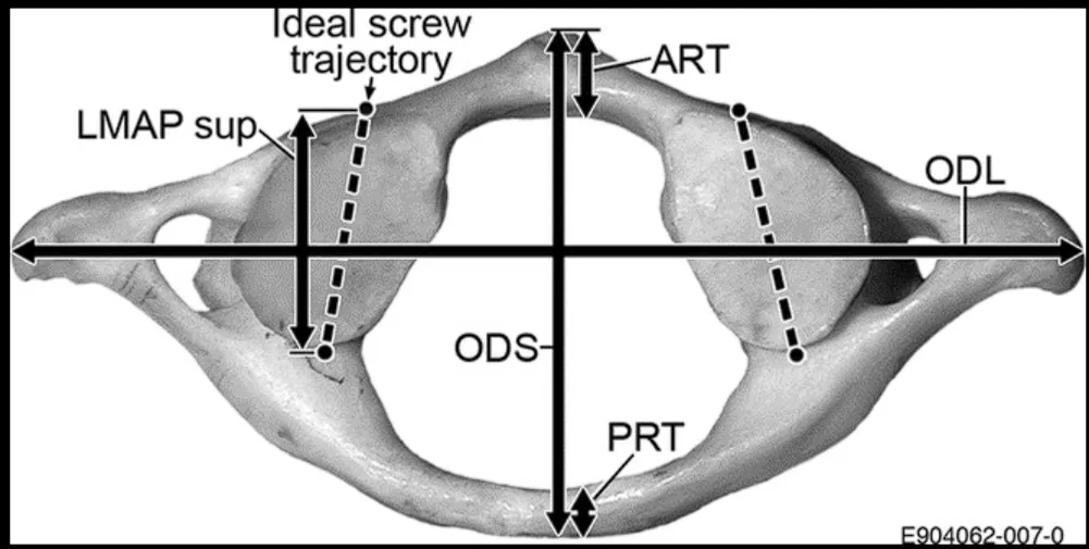
- Superior view of C1 demonstrating the ideal orientation of the screws within the transverse plane (5°–10° medially directed from the sagittal plane), in addition to various other measured dimensions:
- LMAPsup, lateral mass anteroposterior dimension superior to the posterior ring insertion;
- LMAPinf, lateral mass anteroposterior dimension inferior to the posterior ring insertion;
- ODS, outer diameter of the vertebra in sagittal orientation;
- ODL, outer diameter of the vertebra in coronal/lateral orientation;
- ART, anterior ring thickness (in anteroposterior dimension);
- PRT, posterior ring thickness.
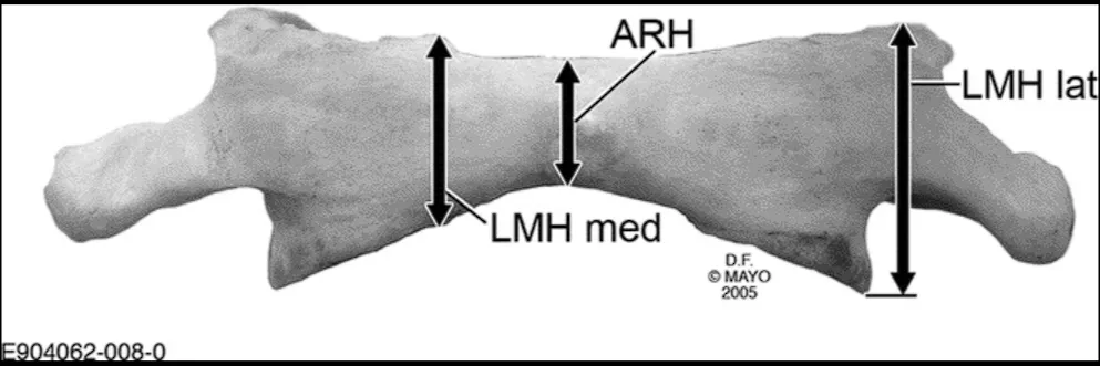
- Anterior view of C1 showing the measurement of anterior height (ARH), as well as the height dimensions of the lateral mass (LMHmed and LMHlat).
 Made with Bullet