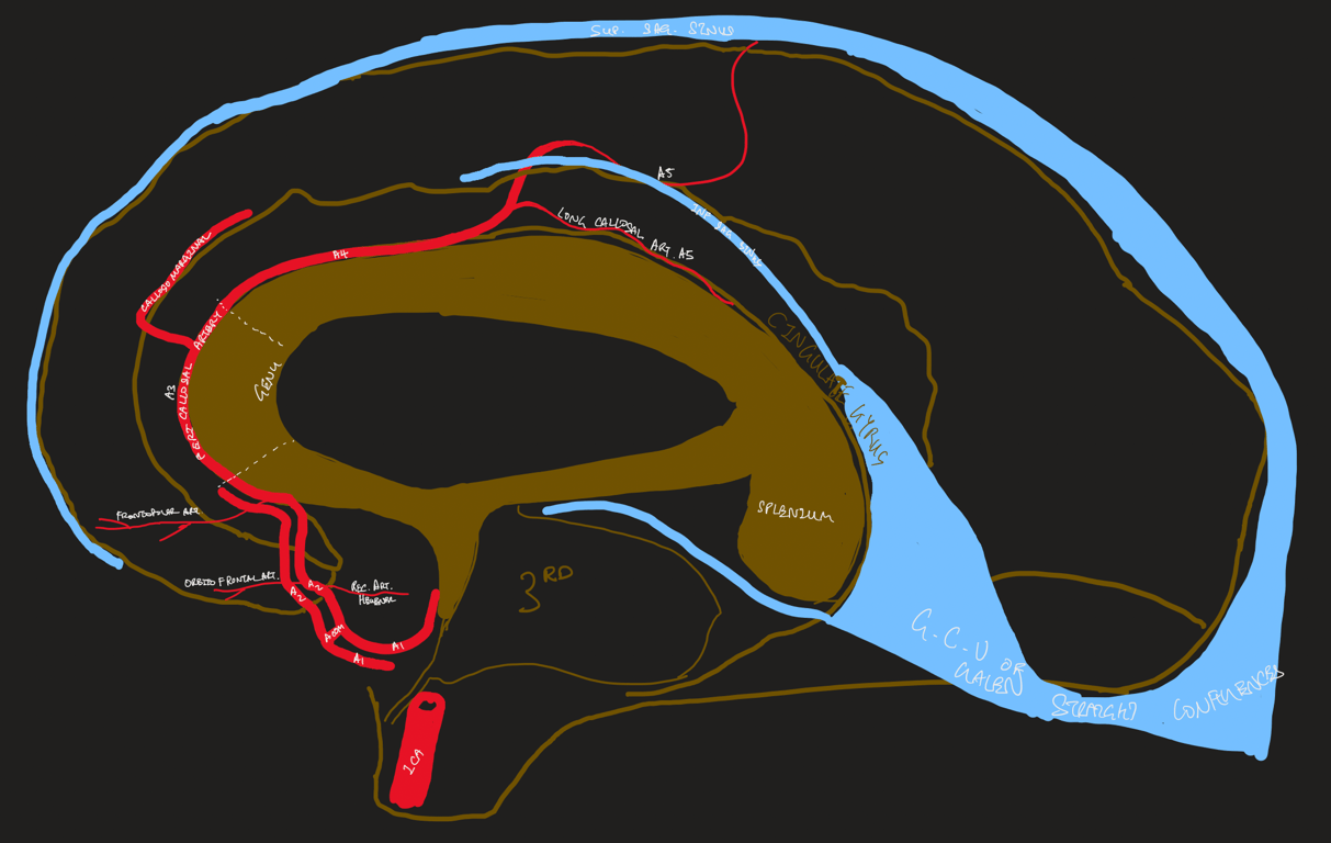
Segments
Horizontal/pre-communicating segment (A1)
- 1-12 perforating arteries: Medial lenticulostriate/medial proximal striate arteries
- Through anterior perforating substance to supply: Optic nerve + chiasm, ant hypothalamus, septum pellucidum , anterior commissure, Pillars of the fornix, Anteroinferior striatum
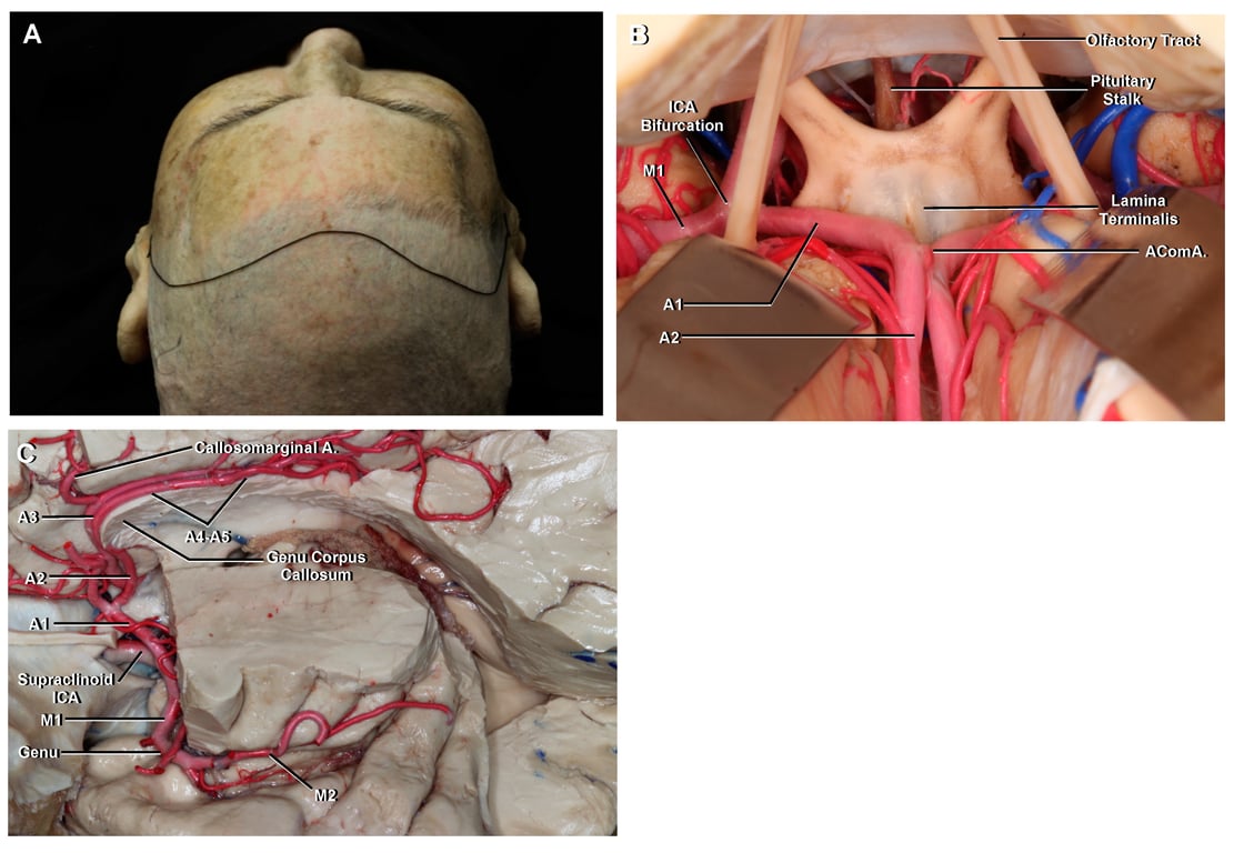
AComA
- Located in cistern of lamina terminalis
- Perforators (3 groups Serizawa classification) Melia 2014
- Hypothalamic branches
- being multiple and of small caliber and ending in the hypothalamic area.
- Chiasmatic branches
- Usually a single vessel and typically the largest of the arteries arising from the AcoA.
- Supplies the
- Bilateral subcallosal areas
- Bilateral columns of the fornix
- Injury: acute confusion due to Korsakoff’s syndrome
- Genu of the corpus callosum
Subcallosal artery (ScA)
- Perforators supply:
- Infundibulum
- Optic chiasm
- Subcallosal area
- Preoptic hypothalamus
- Aneurysm arises at bifurcation of ACOM and A1
- Points towards opposite direction
Vertical/post-communicating segment (A2): Below corpus callosum
H, Recurrent artery of Heubner
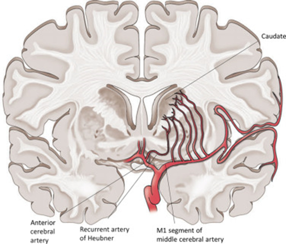
- AKA medical distal striate artery
- This artery is the largest of the perforating branches of the ACA
- Course generally follows the A1 laterally
- Arises anywhere near A1-Acom-A2 J(x): most common @ proximal A2
- Enters anterior perforating substance
- Supplies:
- Head of caudate
- Anterior limb of internal capsule
- Anterior putamen + Globus pallidus
- Septal nuclei
- Inferior frontal lobe
- Easily clipped accidentally when targeting AComA → pure motor stroke
- Unilateral
- Weakness contralateral arm
- Weakness contralateral face
- Dysarthria
- Hemichorea
- Bilateral: akinetic mutism
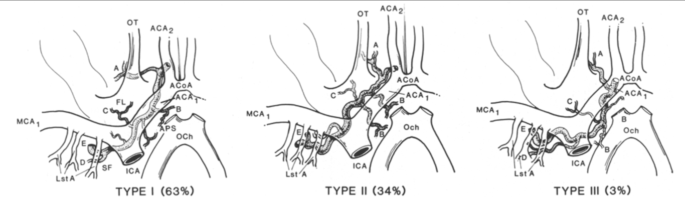
- 3 variation in the course
- (A); anterior perforated substance (APS) branches (B); frontal branches (C); Sylvian fissure branches (D); and terminal branches (E).
- ICA = internal carotid artery; MCA~ = proximal middle cerebral artery; ACA~ = proximal anterior cerebral artery; ACA2 = distal anterior cerebral artery; ACoA = anterior communicating artery; FL = frontal lobe; SF = Sylvian fissure; OT = olfactory tract; Lst A = lenticulostriated arteries; OCh = optic chiasm.
Orbitofrontal arteries
Frontopolar arteries
Distal ACA branches (A3): around level of genu corpus callosum
Callosomarginal artery
- 2nd most common site for anterior cerebral artery aneurysm
- present in approximately 50% of cases
- Supplies the
- superior frontal gyrus through various branches, taking a course through the cingulate sulcus.
- It terminates as the paracentral artery supplying the paracentral lobule
Pericallosal artery/trunk
- Its branches supply the
- corpus callosum and its splenium,
- septum pellucidum,
- fornix,
- precuneus cortex.
- The cortical branches anastomose with the branches of the MCA and posterior cerebral artery
Cortical branches (A4/A5): Above corpus callosum
Anatomical variations
- Unilateral hypoplastic Al segment
- Usually associated with an ACOM aneurysm
- 2 or 3 ACOMs are present in 40% of cases
- Duplicated A1
- If an A1 is hypoplastic, the recurrent artery could be large
- Azygos ACA: the 2 ACAs form a single trunk, and there is no ACOM (present in 0.4 — 1% of the population)
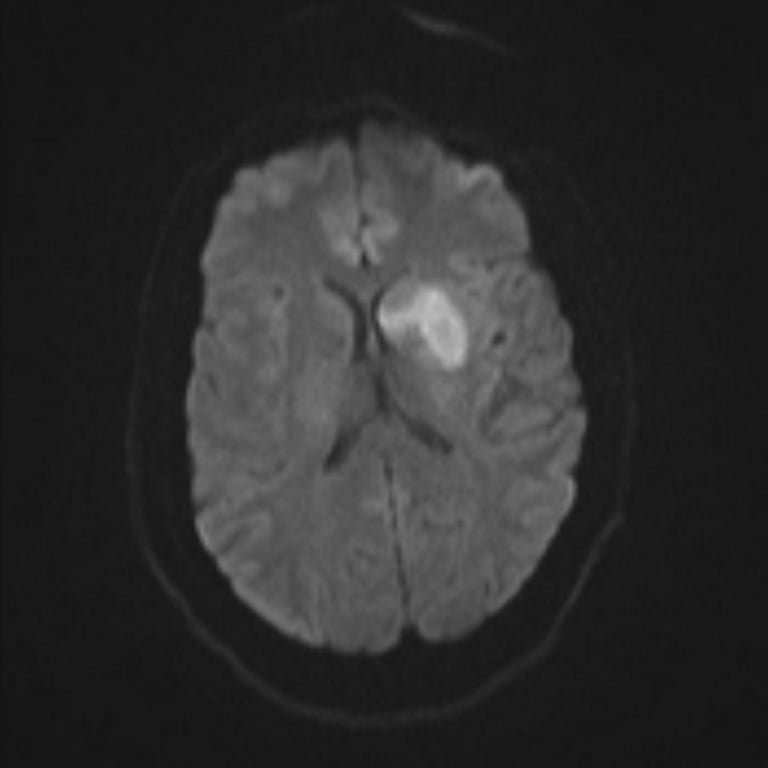
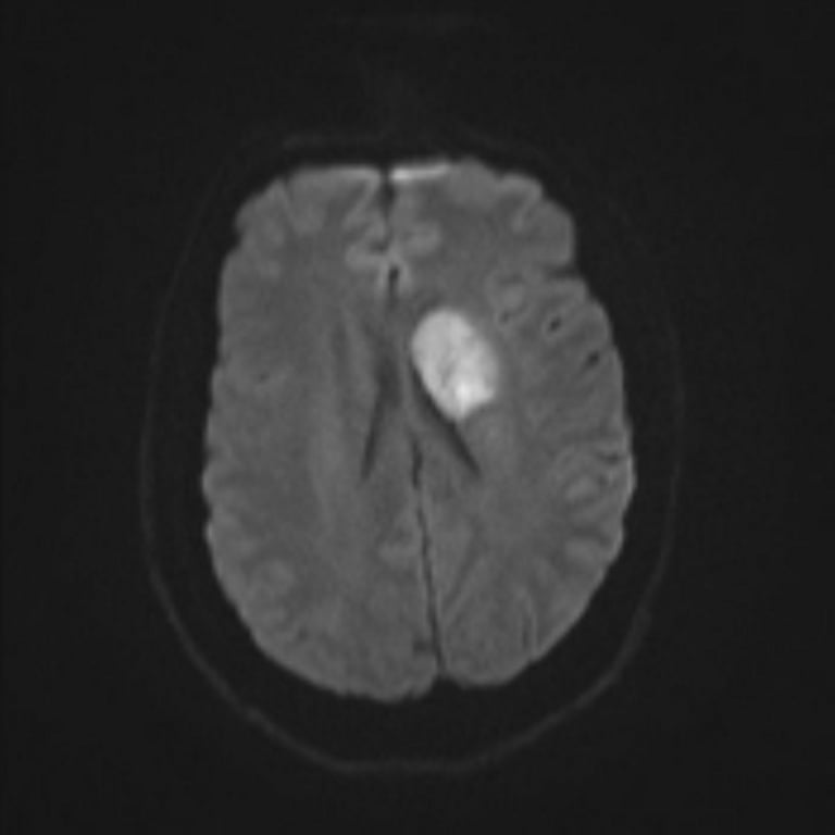
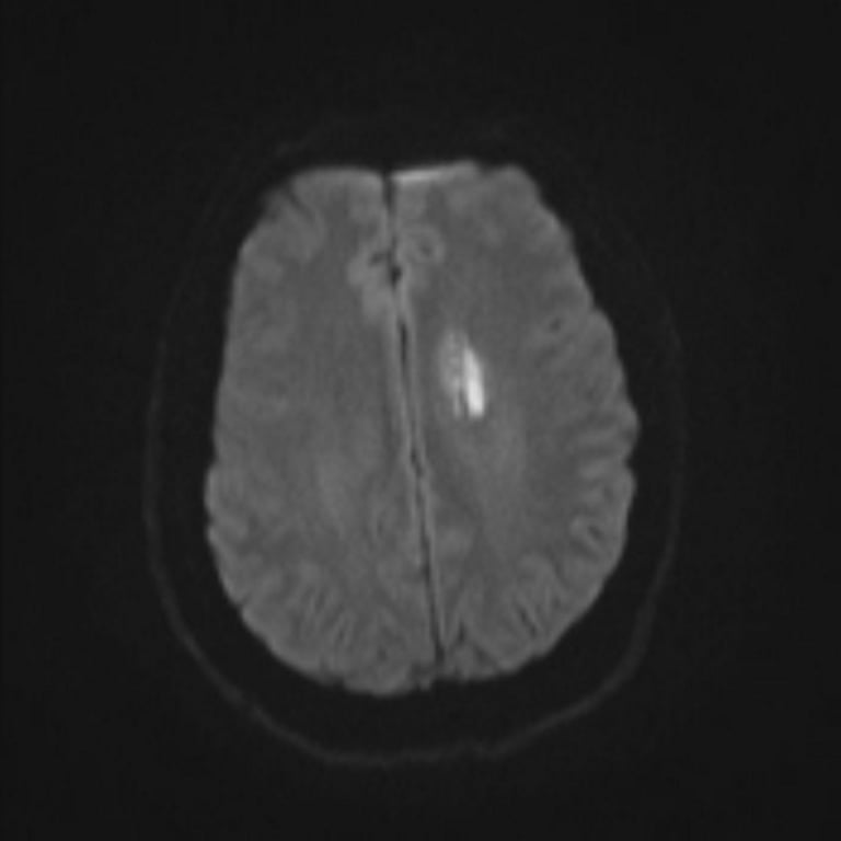
Images
 Made with Bullet