Subclavian artery
- Source
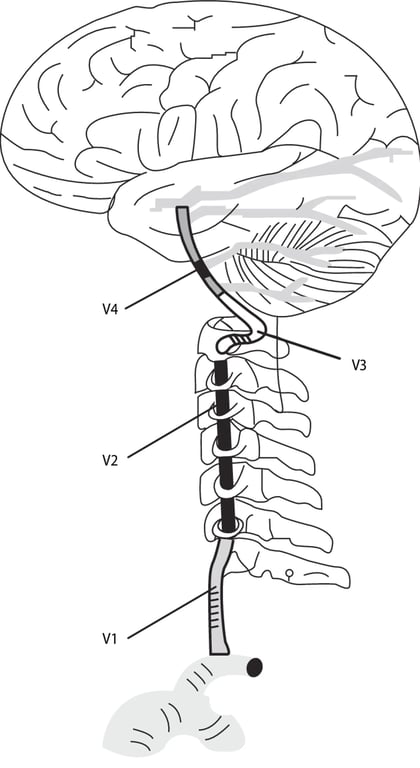
Vertebral artery V1
- Mobile
- Separation: @ C6 enter transverse foramen
Vertebral artery V2
- Immobile
- Separation: Swing behind lateral mass of C1
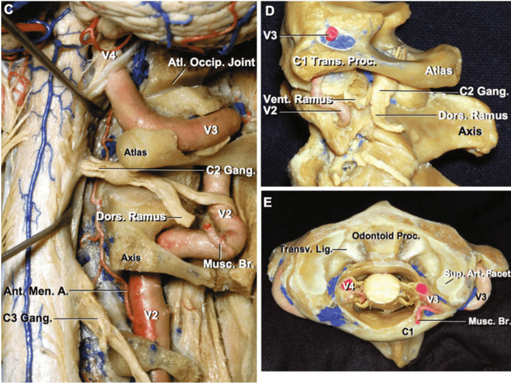
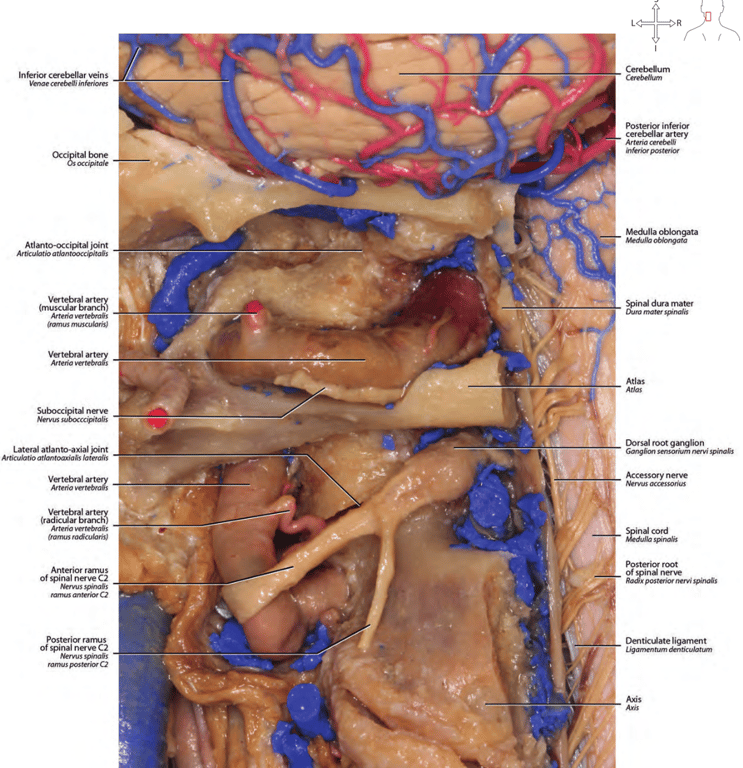
Vertebral artery V3
- Mobile
- Has 3 portions
- Vertical portion:
- ascends through the transverse processes of C1
- Horizontal portion:
- courses in the groove on the upper surface of the posterior arch of the atlas
- Oblique portion:
- penetrates the dura mater
- The distance between the posterior midline and the site of the dural entrance is approximately 12 mm
- Separation: Enter dura
Vertebral artery V4
- Immobile
Posterior spinal artery
- Posterior to dentate ligament
- 1st branch
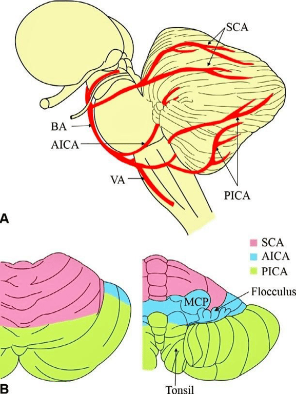
PICA
- Arise intradurally near inferior olive
- 2nd branch
- Largest branch
- sometimes arise below the foramen magnum (~15% individuals)
- PICA courses around the medulla from its origin where it is closely related to the hypoglossal nerve passing around the inferior aspect of the medullary olive—termed the anterior medullary segment
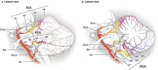
Segments (and separators)
Separation: CN12
Separation: CN 9/10/11
Separation: Medial tonsil midpoint
(Telo: branches to 4th ventricle choroid plexus-tela choroidea; Velo: Roof of 4th ventricle-inferior medullary vellum)-distal from P4 is safer to get Occlusion or sacrificed. Also called choroidal point as the start of this segment is the level of the 4th ventricle. Used in the past when no cross sectional imaging to check for shift in 4th ventricle.
Separation: Tonsillo-biventral fissure
- Medial trunk
- Supplies vermis and adjacent part of hemisphere
- Lateral trunk
- Supplies cortical surface of tonsil and hemisphere
Disease associated with PICA
- Wallenberg (lateral medullary) syndrome
- Tumours
- Meningiomas blood supply
- Petroclival meningiomas
- Branch of meningohypophyseal trunk (ICA)
- Subarcuate artery branch AICA
- Medial clival artery br. Of cavernous carotid
- Jugular branch of occipital artery
- Jugular branch of ascending pharyngeal artery
- Foramen magnum meningiomas
- Branches from V3 segment (anterior meningeal artery
- Posterior meningeal artery branch of VA if posteriorly located
- Hypoglossal branch of occipital artery
- Hypoglossal branch of ascending pharyngeal artery
- Aneurysms
Anterior spinal artery
- last branch
- Separation:
- Passes posterior to dorsal + ventral roots of C1
- Passes ant to dentate ligaments and spinal portion of CN11
- V4 unions at interpeduncular cistern to form basilar artery
Basilar artery
- Immobile
- Branches of Basilar artery from caudal to rostral:
AICA
- Arise close to CN 6
- Between
- CN9
- CN7/8
Segments
- A1: Anterior pontine: inf. Olive
- A2: Lateral pontine: floccule
- Branches into
- Labyrinthine artery
- Branches into subarcuate artery
- Puncture dura @ subarcuate fossa to enter subarcuate canal
- Supplies semicircular canal and petrous bone
- A route of entry from middle ear to brain for infection
- Segments (meatal in the sense of IEM)
- Premeatal segments
- Postmeatal segments
- A3: Flocculopeduncular: Middle cerebellar peduncle
- Bifurcates (near the facial-vestibulocochlear nerve complex) into
- Rostral trunk passes above the flocculus to course on the middle cerebellar peduncle,
- Caudal trunk supplies the area below the flocculus
- A4: Cortical
Paramedian artery
- Supplies
- Ventral pons
- Midbrain
Pontine arteries
- Supplies ventral pons
SCA
- Most constant infratentorial artery
- Arise below the CN3
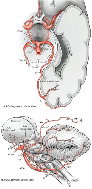
Segments
- Rostral: supplies vermis
- S1: Anterior pontomesencephalic:
- Starts below CN3
- CN5 start at pons
- S2: Lateral pontomesencephalic: Ant margin of Cerebellomesencephalic fissure
- Caudal
- S3: Cerebellomesencephalic: enters cerebellomesencephalic fissure
- S4: Cortical
Supplies:
- Deep cerebellar nuclei (dentate nucleus
- whole superior surface of the cerebellar hemispheres down to the great horizontal fissure
- superior vermis
- most of the cerebellar white matter
- parts of the midbrain
- superior and middle cerebellar peduncle
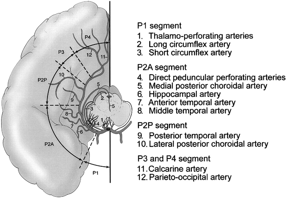
P1 pre-communicating PCA
- Immobile
Branches
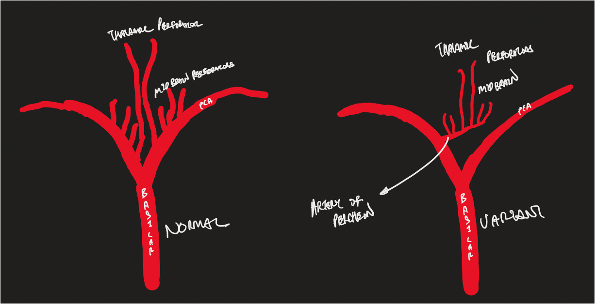
Posterior thalmoperforating arteries: a direct perforating artery
Supply:
- Anterior and part of the posterior thalamus and hypothalamus,
- Somatesthetic disturbances
- Memory deficits: hypothalamic tracts enter/exit between hypothalamus and mamillary bodies
- Autonomic imbalance
- Abnormal movements: cerebellothalamic circuits in the midbrain and thalamus
- Endocrine disturbances: hypothalamic-pituitary axis
- Subthalamus
- Contralateral hemiballismus, which abates into choreiform movements with time or treatment
- Medial part of the upper midbrain: (substantia nigra, red nucleus, oculomotor and trochlear nuclei)
- Diplopia
- Abnormal movements: cerebellothalamic circuits in the midbrain and thalamus
- Oculomotor nerve
- Diplopia
- Mesencephalic reticular formation
- Alterations of consciousness
- Pretectum
- Rostromedial floor of the fourth ventricle
- Posterior portion of the internal capsule
- Motor weakness
- Syndrome will depend on the size of the ischaemia
- Weber syndrome
- Thru' the posterior perforated substance into brain
- Variation: Artery of percheron
- Blockage of this vessel causes
- Bilateral paramedian thalamic stroke
- Vertical gaze palsy
- Memory impairment
- Coma
- Mesencephalothalamic syndrome
- S&S of bilateral paramedian thalamic stroke AND
- Oculomotor disturbance
- Hemiplegia
- Cerebral ataxia
- Movement disorder
Circumflex arteries (circumflexing around the brain stem)
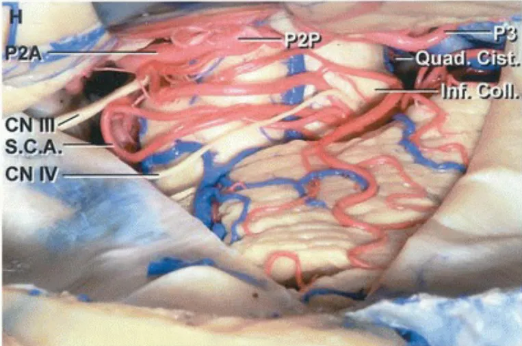
- Long circumflex arteries (quadrigeminal arteries)
- reaches the colliculi
- Supply: quadrigeminal bodies
- Superior colliculus is supplied by Long circumflex artery
- Thrombus: Parinaud's syndrome
- Vertical gaze palsy caused by infarction of the posterior commissure or the nuclei of Cajal
- Inferior colliculus is supplied by branches of the SCA
- Anastomose: with branches from the superior cerebellar artery
- Short circumflex arteries
- reaches the geniculate bodies
- Supply cerebral peduncle
Branch to the quadrigeminal plate
Meningeal branches
Variations
- Absent/hypoplastic P1 segment -
- PCA takes its origin directly from the ICA via a large PCOM
- Foetal origin of the PCA
- 20%
- Large P1
- could be associated with absent/hypoplastic PCA
- PCA duplication
P2 Post communicating PCA
- Immobile
P2A (Anterior @ Crural cistern)
Branches
- Hippocampal artery
- Supplies hippocampal formation
- Enter hippocampus by penetrating the
- Dentate gyrus
- Fimbriodentate sulcus
- Hippocampal sulcus
- Anterior temporal artery
- Supplies the anteroinferior surface of the temporal lobe
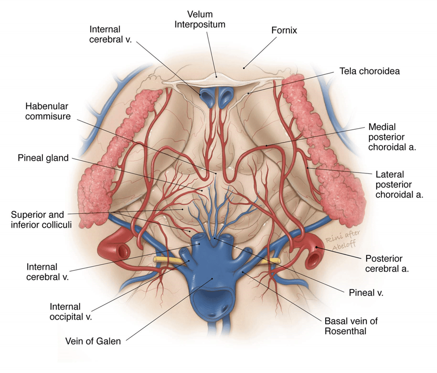
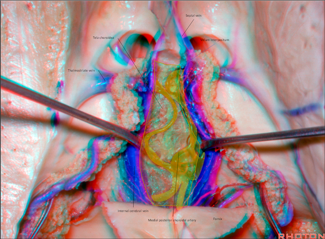
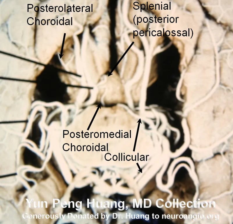
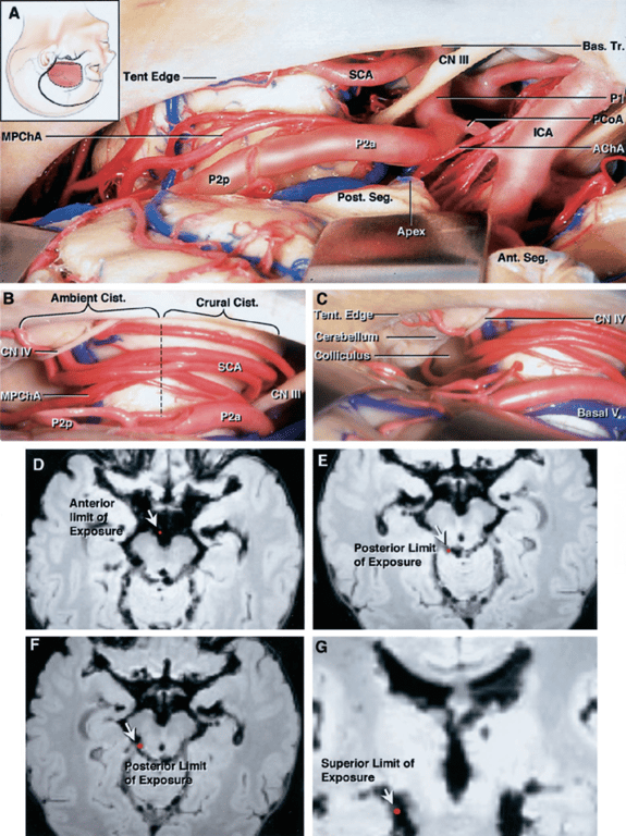
Medial posterior choroidal artery (MPChAs)
- Origin:
- They arise most frequently from the posteromedial aspect of the proximal part of the PCA (P1 or P2) in the interpeduncular and crural cisterns.
- Course
- Travels inferior and medial to the PCA through the crural → ambient cisterns and turns medially to enter the quadrigeminal cistern.
- Passes beneath the splenium of the corpus callosum
- The artery then turns forward to enter the velum interpositum and supplies the choroid plexus in the roof of the third ventricle
- Destination: Lateral + 3rd ventricle
- Supplies:
- Cerebral peduncle
- Tegmentum
- Geniculate bodies (medial > lateral)
- Colliculi
- Pulvinar
- Pineal gland
- Medial thalamus
- Thalmolgeniculate branches: A direct perforating artery
- Peduncular perforating artery: A direct perforating artery
- Supply
- Corticospinal tract
- Corticobulbar tract
- Substantia nigra
- Red nucleus
- Tegmentum
- +/- Oculomotor nerve
- Clinical importance
- P2 branches to the temporal and parietal lobes have relevance to arteriovenous malformations (AVM) surgery.
- The temporal-occipital area receives blood
- Inferiorly from the posterior inferior temporal artery and
- Anteriorly from branches of the MCA.
- P2, in the presence of high intracranial pressure, can be compressed against the tentorium resulting in occipital lobe infarcts.
P2P (Posterior @ Ambient cistern)
- Branches
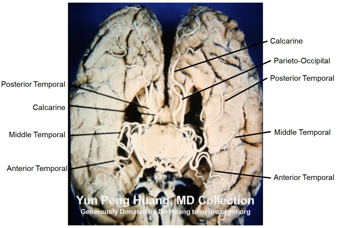
- Middle temporal artery
- Smallest and fq absent
- Supplies inferior surface of the temporal lobe
- Posterior temporal artery
- Largest
- Supplies
- inferior temporal
- Occipital surfaces,
- Occipital pole
- Lingual gyrus
- Occlusions:
- Dysphasia
- Amnestic syndrome
- Homonymous hemianopias
- Colour Agnosia: Inability to match colours to their names
- without hemiparesis or sensory loss
- Common temporal artery
- Supplies:
- Majority of the inferior surface of the temporal and occipital lobes.
- Origin: They arise from the PCA (most commonly the P2P segment) or its cortical branches in the ambient and quadrigeminal cisterns.
- Course
- They pass laterally around the pulvinar
- Course laterally along the upper edge of the parahippocampal gyrus within the ambient cistern
- Pass through the choroidal fissure to enter the posterior part of the temporal horn and atrium
- enters the ventricle adjacent to the lateral geniculate nucleus through the choroid fissure
- Destination: temporal horn of lateral ventricle
- The number of LPChAs in a hemisphere averages four (ranging from one to nine).
- Supplies:
- Cerebral peduncle
- Posterior commissure
- Fornix (Part of crura and body)
- Lateral geniculate body
- Pulvinar
- Dorsomedial thalamic nucleus
- Body of the caudate nucleus
- Anastomosis with
- Ant Choroidal Artery
- Medial Posterior Choroidal Artery
Lateral posterior choroidal artery (LPChAs)
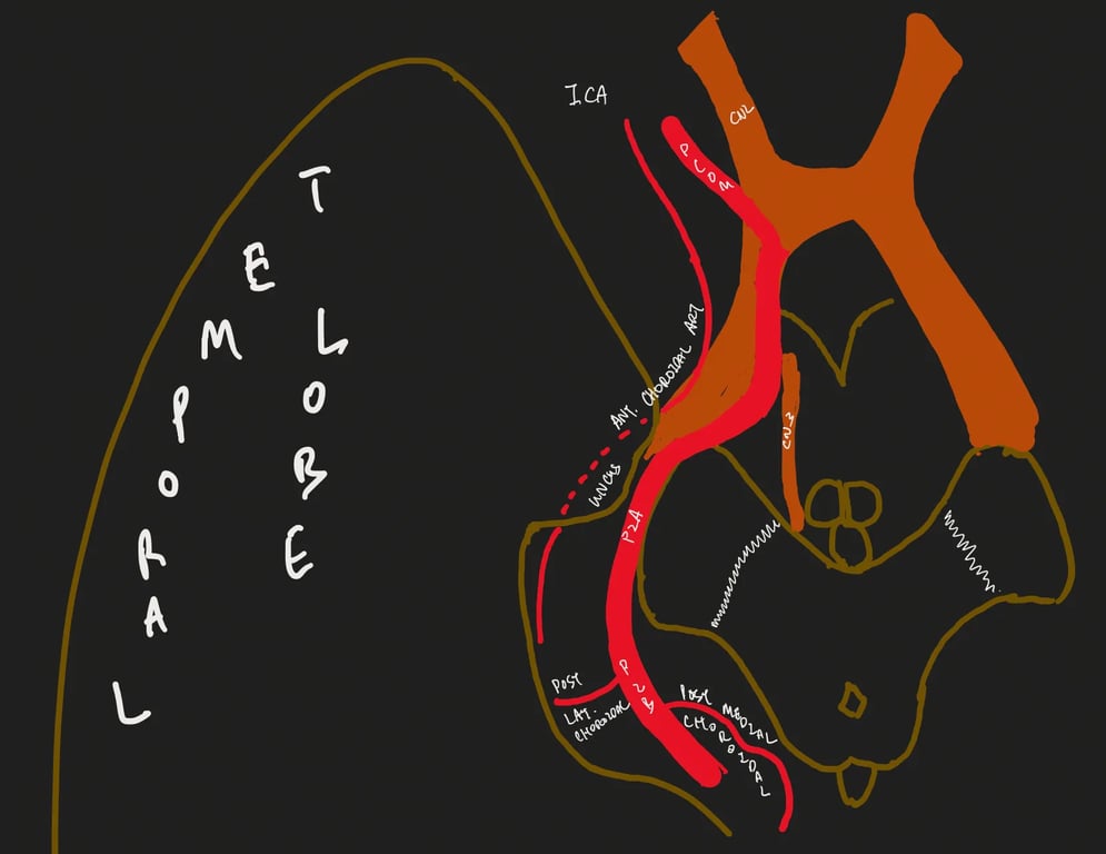
P3 PCA
- @ Quaddrigeminal cistern
- Immobile
- ends at calcarine fissure
- Collicular or quadrigeminal point: when bilateral P3 comes closest to each other (8.9 mm apart)
P4 PCA
- @ quadrigemial cistern
- Cortical branches
Inferior temporal arteries (superior temporal comes from MCA)
- inferior temporal branches
- See P2A
- Anterior temporal branches
- See P2P
- Common temporal artery
- Middle temporal branches
- Posterior temporal branches
- Hippocampal arteries
- Supplies
- Uncus
- Anterior parahippocampal gyrus
- Hippocampal formation
- Dentate gyrus
- Bilateral occlusion: syndrome resembling korsakoff syndrome (severe memory loss)
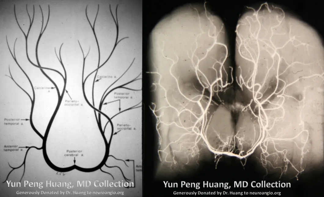
Internal occipital artery
- Calcarine artery @calcarine sulcus
- Supply
- Lingual gyrus (medial occipitotemporal gyrus)
- Inferior cuneus
- visual cortex
- Occipitoparietal artery @occipitoparietal sulcus
- Supply
- Posterior parasagittal region
- Cuneus
- Precuneus
- Lateral occipital gyrus
- Occlusion
- Unilateral
- Homonymous visual field defect with macular sparing
- pain in the ipsilateral eye
- Bilateral
- blindness with preserved pupillary reflexes OR
- Anton’s syndrome
- A form of anosognosia restricted to vision
- Preservation of the pupillary reaction to light
- Patient denies there is any visual disturbance despite being functionally blind.
- Patient will confabulate
- Damage:
- Primary visual cortex.
- visual field may recover after ligation or occlusion of the calcarine artery
- Posterior pericallosal artery
- Splenial arteries
- Aka Posterior pericallosal artery
- Variable origin: parieto-occipital, calcarine, medial posterior choroidal, posterior temporal, and lateral posterior choroidal
- anastomose with branches of the pericallosal artery
- Occlusion → splenium of the corpus callosum ischaemia → interrupts the fibers between the intact occipital pole and contralateral angular gyrus → syndrome of dyslexia (difficulty reading) without dysgraphia (difficulties with spelling, poor handwriting and trouble putting thoughts on paper)
Images
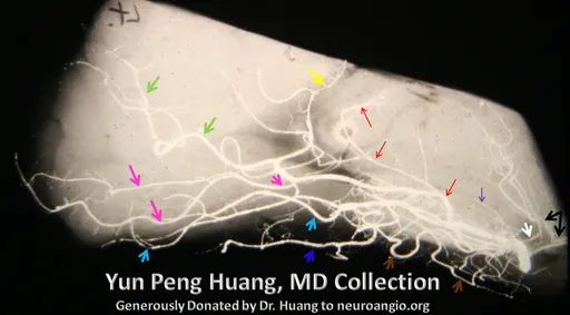
Black: PCOM
Purple: Geniculate
Red: Posterior lateral choroidal (note characteristic C-shaped curve)
Posterior pericallosal (a.k.a. splenial branch) = yellow
Calcarine = pink
Parieto-occipital = green
Posterior Inferior Temporal = light blue
Middle Inferior Temporal = dark blue
Anterior Inferior Temporal = brown
 Made with Bullet