General
- Single ventral arterial axis
- Anterior spinal artery (ASA)
- The anterior axis lies within the ventral spinal cleft
- forms from a series of radiculomedullary arteries, while the posterior form from radiculopial vessels.
- Two dorsal arterial axes— the posterior spinal arteries (PSA).
- Branches of the radicular arteries reach the cord by following the dorsal and ventral roots.
- Two “watershed” areas within the anterior spinal artery at the level of T1-T3 and T6-T8.
- These are regions that lie in between the three main vascular origins, thereby receiving arterial blood both from above and from below; the resulting oppositional flow can lead to cord hypo-perfusion at lower blood pressures.
- Due to Caton 2023
- less robust anastomoses exists between T4 and T8, rendering these levels susceptible to transient perfusion abnormalities
- But clinically not entirely proven
- Some explain relative hypovascularity of the mid-thoracic cord reflects metabolic parsimony due to relatively decreased oxygen demand at these levels relative to other cord regions
Spinal artery embryogenesis
- These ventral and dorsal axes themselves develop in- utero as paired metameric arteries.
- 4-6th week gestation:
- Fusion
- of the anterior metameric pairs ultimately → ASA.
- Failure of fusion accounts for the occasional fenestrations seen in the ASA.
- Desegmentation: occurs with involution of many metameric arteries.
- Simultaneously occurs with fusion
- 62 metameric vessels initially
- dorsal 31 → 10-20 remain
- ventral 31 → 4– 8 remain
- There is great variability in the degree to which anterior metameric arteries regress, but two relatively constant remnants are named.
- The artery of the lumbar enlargement
- Aka
- Larger radiculomedullary artery
- Arteria radicularis magna
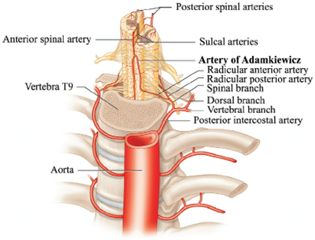
- Artery of Adamkiewicz
- Most often arises from the left side.
- 75% of cases it arises between T9 and T12.
- When it arises outside of that area there is usually a second large, persisting metameric artery to be found cranially or caudally.
- Artery of Adamkiewicz.
- In 75% of patients, the AKA arises between T9 and T12, more commonly on the left.
- When its origin is above T8 or below L2, another major contributor to the ASA can be found either cranially or caudally.
- In 30-50% of cases, it also contributes significantly to the PSA.
- Generally, a pair of arteries arises in the cervical region from the intradural segment of each vertebral artery that fuse to one “Y”-shaped ASA running in the subpial space in the ventral sulcus of the spinal cord (dorsal to the anterior spinal vein) to the terminal film.
- The typical hairpin anastomosis between the radiculomedullary arteries and the ASA is found angiographically at the lower thoracic and lumbar levels.
- The artery of the cervical enlargement
- Entering the C5/ 6 level
- arises from
- Thyrocervical trunks
- Costocervical trunks.
- subclavian artery (rarer)
- Variation is again frequent
- 15– 20 weeks gestation
- Posterior axes fuses
The radicular arteries
- Radicular artery
- Radiculospinal branches. (Neuroangio does not talk about radiculospinal arteries instead radicular artery directly becomes radiculopial or radiculomedullary)
- Supplies the spinal cord
- At each level, the radiculospinal branch may divide into → entering the spinal canal with the corresponding nerve roots
- anterior branches
- posterior branches
- This extrinsic arterial supply is relatively well collateralized, particularly in the cervical region.
- Occlusion of the costocervical artery is relatively unlikely to result in anterior spinal infarction, as the deep and ascending cervical arteries will collateralize the anterior spinal axis.
- Clinical
- Radiculospinal branches form Spinal AVM and perimedullary AVF
- Arriving at the cord each radiculospinal artery can be formed into two variants
- Radiculopial arteries
- Supplies the posterior axis
- The terminal branches of the radicular arteries, reaching the pia via the spinal root, form PSA.
- Those branches reaching the cord are termed radiculopial arteries and they invade the cord from the pial surface and supply the peripheral parts of the cord with a watershed region at the interface between the white and grey matter.
- Vasa coronae.
- Small branches from radiculopial and radiculomedullary systems run on the pial surface and encircle the cord.
- These give perforating vessels into the superficial white matter of the cord as well as forming a superficial anastomosis with the anterior circulation.
- Radiculomedullary artery
- An enlarged radicular artery instead of supplying local neural elements, it maintained its embryonic access to the anterior spinal artery.
- Supplies the anterior axis
- Most of the anterior grey matter of the spinal cord
- Anterior part of the posterior grey matter
- Elements of the anterior, lateral, and posterior white columns.
- Each radiculomedullary artery contributes a superior and inferior ramus to form the anterior arterial axis.
- Sulco-commissural arteries penetrate the cord via the ventral commissure in turn giving rise to the radially directed perforating vessels, which supply the cord.
- Radiculomeningeal branches.
- Supply the nerve root’s dural sheath and contents
- Do not supply the spinal cord.
- Clinical
- Radiculomeningeal branches form arterial supply for Spinal dural AV fistulae
Arterial anastomoses and the ‘conus basket’
- The vasa coronae and radial perforating vessels form longitudinal and segmental anastomoses along the cord.
- The relative contributions by the radiculopial and radiculomedullary systems vary.
- Cervical cord is evenly served
- Thoracic cord
- Relatively less reliably served
- Although the well- known vulnerability of the thoracic cord to ischaemia may also be a function of its venous drainage as discussed next.
- Lumbosacral Cord
- rely mainly on the central radiculomedullary system
- Conus basket
- An especially well-developed anastomotic channel between anterior and posterior arterial axes
- The ASA tapers as it runs down the anterior surface of the conus and one to two branches join it to the PSA (Martirosyan et al., 2015).
- At the level of the conus medullaris.
- The artery of the filum terminale is the terminal continuation of the anterior axis below the conus.
Normal arterial supply (Neuroangio)
- Supply tree for spinal cord
flowchart TD
    A["Aorta (A)"] --> B["Segmental artery (B)"]
    B --> E["Dorsal spinal artery (E)"]
    E --> K["Ventral division of dorsal<br>spinal artery (K)"]
    K --> Ka["Radicular artery (Ka)"]
    Ka --> O["Radiculopial artery (O)"] & P["Radiculomedullary artery (P)"]
    O --> X["Rami perforantes of<br>the peripheral system (X)"] & S["Posterior spinal<br>artery (S, T)"]
    P --> Q["Anterior spinal artery (Q)"]
    Q --> W["Sulco-commissural<br>artery (W)"]
    W --> Y["Central system of<br>sulcal arteries (Y)"]
- Supply tree for meninges
flowchart TD
    A["Aorta (A)"] --> B["Segmental artery (B)"]
    B --> E["Dorsal spinal artery (E)"]
    E --> K["Ventral division of dorsal spinal artery (K)"]
    K --> Ka["Radicular artery (Ka)"]
    Ka --> n1["Epidural fat supply"] & n2["Theca supply"]
    M["Nerve root sleeve<br>dural branch of the<br>ventral division<br>dorsal spinal artery (M)"] --> N["Dural branch of the<br>ventral division<br>dorsal spinal artery (N)"]
    n2 --> M
    n1 --> Lb["Dorsal<br>epidural arcade (Lb)"] & La["Ventral<br>epidural arcade (La)"]

    n1@{ shape: hex}
    n2@{ shape: hex}
    style n1 fill:#FFD600,color:#FFFFFF
    style n2 fill:#2962FF,color:#FFFFFF
Images
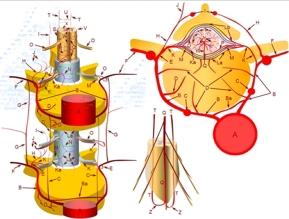
A – aorta | B – segmental artery | Ba – intersegmental arterial anastomosis |
C – prevertebral anastomotic network | D – direct vertebral body feeding arteries | E – dorsal spinal artery |
F – intercostal/muscular artery | G – pretransverse anastomotic network | H – dorsal division of the dorsal spinal artery |
I – post-transverse anastomotic network | J – muscular branches of the post-transverse anastomotic network | K – ventral division of the dorsal spinal artery; |
Ka – radicular artery | La – ventral epidural arcade | Lb – dorsal epidural arcade; |
M – nerve root sleeve dural branch of the ventral division dorsal spinal artery | N – dural branch of the ventral division dorsal spinal artery | O – radiculopial artery; |
P – radiculomedullary artery | Q – anterior spinal artery | R – mesh-like pial arterial network |
S, T – posterior spinal artery; | U, V – pial arterial network (a.k.a. vasocorona) anastomoses between anterior and posterior spinal arterial systems, | W – sulco-commissural artery, |
X – rami perforantes of the peripheral (centripetal) system, | Y – central (centrifugal) system of sulcal arteries, originating from pial network of the cord; altogether, the pial network and rami perforantes (R+Y) are called the vasocorona or corona vasorum; | Z – rami cruciantes (a.k.a. crux vasculosa, a.k.a. rami anastomotici arcuati) |
 Made with Bullet