General
- Situated lateral to the sella, resting on the sphenoid and temporal bones
- A paired venous structure
Enclosed by five dural walls
- These dural folds that envelop the cavernous sinus and its contents serve as consistent landmarks and provide routes for surgical access to the cavernous sinus
Laterally
- Faces the temporal lobe laterally
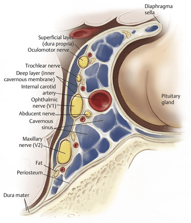
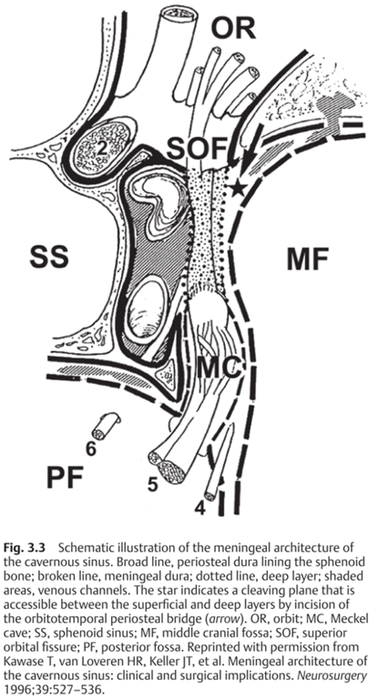
- The dura of the middle fossa is composed of an outer periosteal layer that is adherent to the inner surface of the cranium, and an inner meningeal layer that faces the brain. At the lateral edge of the cavernous sinus, the middle fossa dural lining separates.
- The meningeal layer turns upward as the temporal lobe dura and forms the superficial layer of the lateral wall of the cavernous sinus
- The periosteal layer continues medially along the skull base to become the medial wall.
- lateral wall of the cavernous sinus reveals a two-layered construct.
- Superficial layer
- Continuation of the middle fossa meningeal dura
- Can be easily separated from a thinner deep layer.
- Deep layer
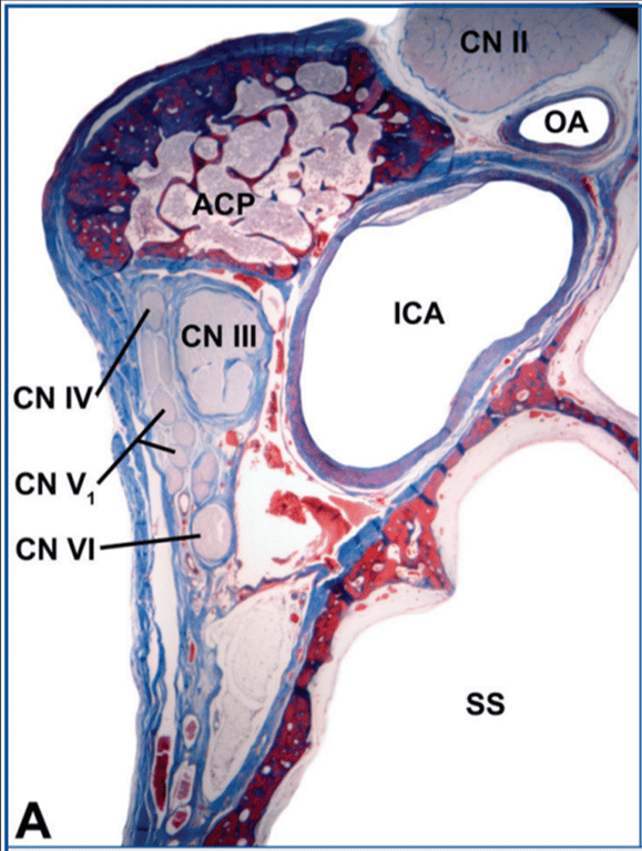
- The dural sheaths that accompany cranial nerves III, IV, V1, and V2 as they penetrate into the sinus form this semitransparent deep layer.
- Superolateral view; the left anterior clinoid has been removed.
- The proximal and distal dural rings in the lateral wall of the carotid artery delimit the clinoid segment of the carotid artery.
- These rings typically fuse at the posterior aspect of the carotid artery medial to the tip of the anterior clinoid process.
- Ligaments of the cavernous sinus, superior view.
- The dura mater forming the superior wall of the left cavernous sinus has been removed along with the posterior half of the distal dural ring to show the interclinoid ligament between the anterior and posterior clinoid processes and the anterior and posterior petroclinoid ligaments between the petrous apex and the anterior and posterior clinoids, respectively.
- The left anterior clinoid has been removed.
- The carotidoculomotor membrane forms the proximal dural ring when it meets the carotid medially.
- Blue silicon, representing the venous filling in the cavernous sinus, has been partially removed below the distal dural ring posteriorly.
- The proximal dural ring is not evident in the posterior and medial aspect of the carotid.
- In some specimens, there is a carotico clinoid ligament between the anterior clinoid and middle clinoid (when present) or medial border of the carotid sulcus at the level of the anterior bend of the carotid artery (when the middle clinoid is absent).
- Further removal of blue silicon of the left cavernous sinus.
- This ligament blends into the carotidoculomotor membrane and is directed to the periosteal layer covering the middle clinoid process or medial aspect of the carotid sulcus.
- It may have adherences to the internal carotid artery and to the meningeal layer of the dura mater covering the pituitary.
- The left half of the pituitary gland has been removed to show the meningeal layer covering the gland and the periosteal layer covering the sellar wall of the sphenoid sinus.
- Both of them form part of the medial wall of the cavernous sinus.
Dural relationships and ligaments in the cavernous sinus
Medially
- Sphenoid bone
- Sella turcica
- Pituitary gland medially
- Truong et al 2018:
- Unlike the dual-layered lateral wall, the medial wall of the cavernous sinus is composed of a single thin layer of dura that cannot be separated.
- This single-layered construction of the medial wall may explain the tendency for pituitary adenomas to invade the cavernous sinus.
- Although a single layer of dura constitutes the medial wall, it can be divided into two parts, each with a different dural origin.
- Parasellar ligaments
- Function:
- They serve to anchor the medial wall of the cavernous sinus (CS) to other walls of the CS and/or to specific segments of the internal carotid artery (ICA).
- Classification (4 Groups):
- Caroticoclinoid ligament:
- Location:
- Spans from the medial wall and the middle clinoid.
- Connections:
- Connects towards the clinoid ICA segment and the anterior clinoid process.
- Prevalence:
- Present in most CSs (97.7%).
- Characteristics:
- It is the strongest and largest ligament.
- Arrangement:
- Typically assembled as a group of ligaments with a fan-like arrangement.
- Superior parasellar (Paras.) ligament:
- Connections:
- Connects the medial wall to the horizontal cavernous ICA and/or the lateral wall of the CS.
- Prevalence:
- Identified in approximately half of the CSs (57.5%).
- Inferior parasellar ligament:
- Connections:
- Bridges the medial wall to the anterior wall of the CS or the anterior surface of the short vertical segment of the cavernous ICA.
- Prevalence:
- Present in most CSs (95%).
- Surgical Note:
- This ligament is the first to be encountered after opening the anterior wall of the CS during an interdural transcavernous approach.
- Posterior parasellar ligament:
- Connections:
- Anchors the medial wall to the short vertical segment of the cavernous ICA and/or the posterior carotid sulcus.
- Prevalence:
- Identified in approximately half of the CSs (45%).
Anteriorly
- the cavernous sinus fills the posterior margin of the superior orbital fissure below the anterior clinoid process, and it extends posteriorly to the petrous apex.
- The lateral and medial walls join anteriorly at the superior orbital fissure (SOF), and the cranial nerves exiting the apex of the cavernous sinus are wrapped in a common meningeal sheath.
- The middle fossa periosteal dura continues through the SOF as the periosteal layer of the periorbita, creating the orbitotemporal periosteal fold.
- Sectioning of this periosteal bridge is the initial step in the extradural elevation of the superficial layer of the lateral wall and a useful step to enhance extradural exposure of the anterior clinoid process for anterior clinoidectomy.
- Posterolateral to the SOF at the site of middle cerebral vein drainage, venous channels are covered by meningeal dura only.
- This absence of a deep inner membrane creates a weak point in the cavernous sinus wall and a potential route of invasion into the cavernous sinus apex from medial sphenoid wing meningiomas.
Posterior wall
- Stretches from the lateral edge of the dorsum sellae to the medial margin of the Meckel cave.
Roof
- Composed of the clinoidal and oculomotor triangles and faces the basal cisterns.
Layers of the cavernous sinus coverings
- A, diagram showing the dura divided into a meningeal layer (orange) and an endosteal layer (green).
- The two layers are tightly adherent in the floor of the middle cranial fossa, but on reaching the upper edge of the second trigeminal division (V2), which is the most inferior limit of the cavernous sinus, they separate into two layers.
- The meningeal layer extends upward to form the outer layer of the lateral wall and roof of the cavernous sinus and the upper layer of the diaphragma sellae.
- The endosteal layer, at the level of the upper border of the maxillary nerve, divides into two layers.
- One layer extends upward to constitute the internal layer of the lateral wall and roof of the cavernous sinus
- The other one adheres to the sphenoid bone, covering the carotid sulcus and the sellar floor.
- From the free edge of the diaphragm, a thin layer of dura extends downward to wrap around but is easily separable from the pituitary gland.
- Our dissections suggest that the meningeal layer forms the sellar part of the medial wall of the cavernous sinus and that the endosteal layer (green layer) forms the sphenoidal part of the medial wall. The meningeal and endosteal layers of dura fuse into a single layer on the sellar floor.
- B, diagram illustrating that it is easy to separate the meningeal layer covering the inferior aspect of the pituitary gland from the endosteal layer covering the bony sellar floor.
- C, diagram illustrating an inferior intercavernous sinus that connects the paired cavernous sinuses.
- These intercavernous sinuses extend across the midline between the meningeal dural layer covering the inferior aspect of the pituitary gland and the endosteal layer covering the osseous sellar floor.
Contents
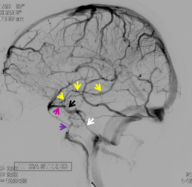
Connections
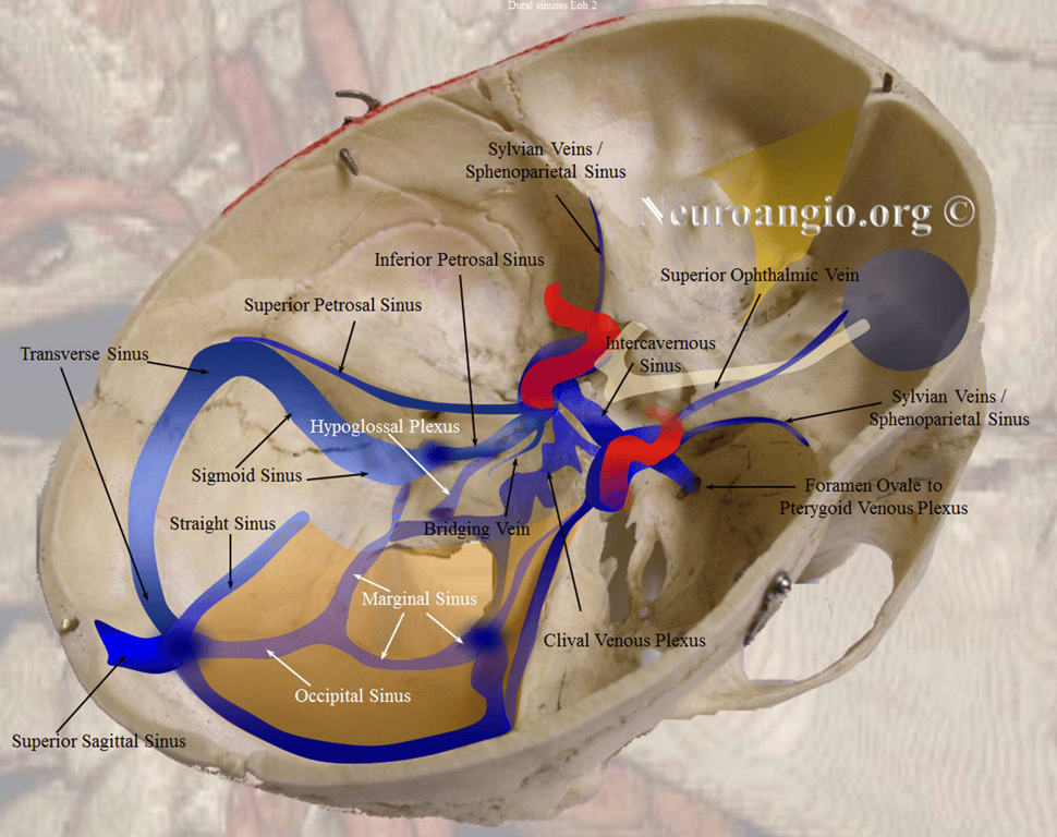
- Inflow
- Superficial middle cerebral sylvian veins (pink)
- Sphenoparietal sinus (itself sometimes draining the Sylvian veins).
- Ophthalmic veins
- The deep sylvian vein – basal vein conduit (yellow, notice full extent of basal vein between CC and the Galen),
- Outflow
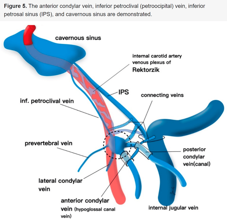
- Inferior petrosal sinus (white)
- Connecting to jugular foramen
- Superior petrosal sinuses
- connecting to sigmoid sinus
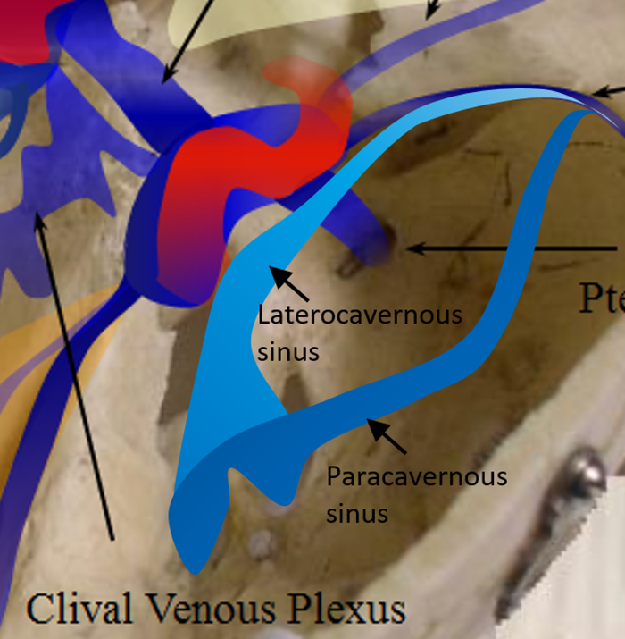
- Pterygoid venous plexus (purple)
- Clival venous plexuses
- Contralateral cavernous sinus
Images
Abbreviation
Abbreviation | Full Form | Abbreviation | Full Form | Abbreviation | Full Form |
A. | Artery | Int. | Internal | Quad. | Quadrigeminal |
A.I.C.A. | Anteroinferior cerebellar artery | Interped. | Interpeduncular | Retrot. | Retrotonsillar |
Ant. | Anterior | Jug. | Jugular | Sag. | Sagittal |
Atr. | Atrial | Lat. | Lateral | S.C.A. | Superior cerebellar artery |
Bas. | Basilar | Lig. | Ligament | Seg. | Segment |
Bivent. | Biventral | Marg. | Marginal | Sig. | Sigmoid |
Br. | Bridging | Med. | Medial, medullary | Str. | Straight |
Carotid | Carotid | Mes. | Mesencephalic | Sulc. | Sulcus |
Cav. | Cavernous | Mid. | Middle | Sup. | Superior |
Cer. | Cerebellar, cerebellum | N. | Nerve | Supracol. | Supracolliculate |
Cer. Med | Cerebellomedullary | Occip. | Occipital | Supraton. | Supratonsillar |
Cer. Mes. | Cerebellomesencephalic | Olf. | Olfactory | Temp. | Temporal |
Cer. Pon. | Cerebellopontine | P.C.A. | Posterior cerebral artery | Tent. | Tentorial |
Ch. | Choroidal | Ped. | Peduncle | Ton. | Tonsillar |
Cist. | Cistern | Pet. | Petrosal | Trans. | Transverse |
CN | Cranial nerve | P.I.C.A. | Posteroinferior cerebellar artery | Trig. | Trigeminal |
Comm. | Communicating | Pon. | Pontine, ponto | V. | Vein |
Condylar | Condylar | Pon. Med. | Pontomedullary | Ve. | Vermian |
Em. | Emissary | Pon. Mes. | Pontomesencephalic | Vel. | Velum |
Fiss. | Fissure | Pon. Trig. | Pontotrigeminal | Vent. | Ventricle |
Hem. | Hemispheric | Post. | Posterior | Vert. | Vertebral |
Inf. | Inferior | ㅤ | ㅤ | ㅤ | ㅤ |
 Made with Bullet