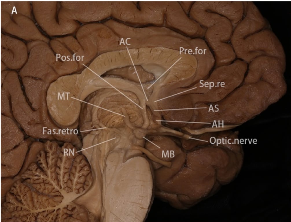
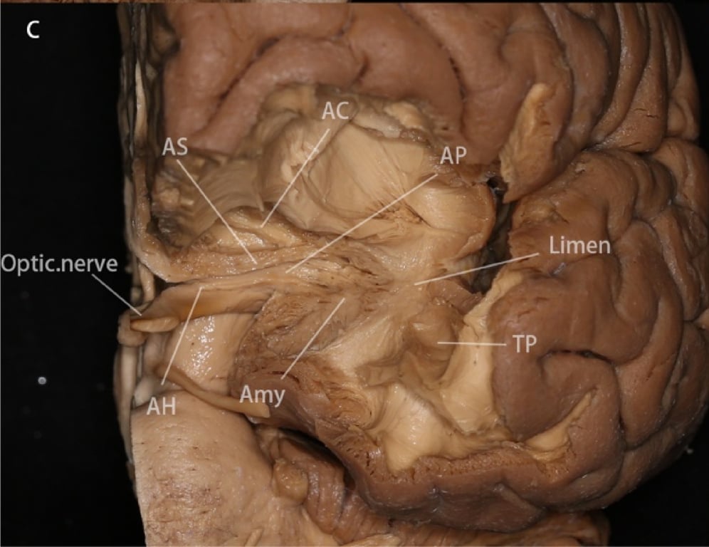
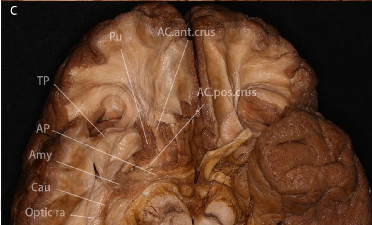
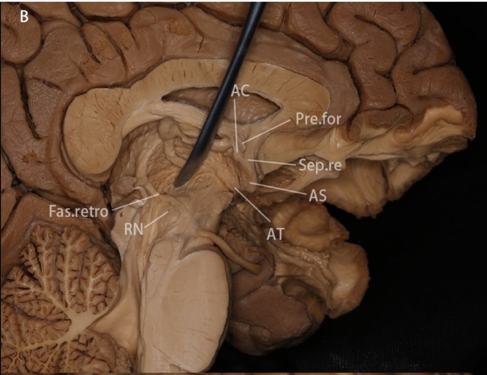
Amygdalofugal pathway

Composed of

Images

Key
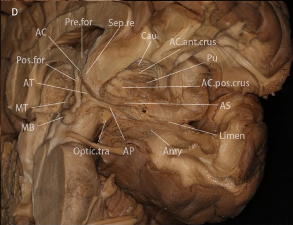
  • AC = anterior commissure
  • AC.ant.crus = anterior crus of the anterior commissure
  • AC.pos.crus = posterior crus of the anterior commissure
  • AH = amygdalohypothalamus
  • Amy = amygdala
  • AP = ansa peduncularis
  • APS = anterior perforated substance
  • AT = amygdalothalamic pathway
  • AS = amygdaloseptal pathway
  • Cau = caudate
  • Fas retro = fasciculus retroflexus
  • GP = globus pallidus
 
  • Hippo = hippocampus
  • Lolfs = lateral olfactory stria
  • MB = mammillary body
  • MT = mammillothalamic tract
  • Optic.tra/Optic ra = optic radiation
  • Pos.for = postcommissural fornix
  • Pre.for = precommissural fornix
  • Pu = putamen
  • RN = red nucleus
  • Sep.re = septal region
  • TP = temporal pole
  • UF = uncinate fasciculus
 
A: Part of the thalamus and septal cortex were removed to expose the precommissural and postcommissural fornix, anterior commissure, mammillothalamic tract, fasciculus retroflexus and amygdaloseptal pathway. The amygdaloseptal pathway is seen arising from the septal region (consisted of the subcallosal cortices and paraterminal gyrus) and running anterior to the body of the anterior commissure, to later turn laterally and form part of the ansa peduncularis. The amygdalohypothalamic fibers originate from the hypothalamus.
C: Anterior view of the ansa peduncularis. The gyrus rectus, orbital gyrus, lateral and media olfactory stria, olfactory tubercle, anterior perforated substances, part of the nucleus amygdala and temporal pole were removed to expose the ansa peduncularis, which extends laterally superior to the optic tract and inferior to the anterior commissure, and reaches the amygdala and anterior temporal cortex.
A: The gyrus rectus, orbital gyrus, occipital-temporal gyrus and parahippocampal gyrus were removed to expose the uncinate
fasciculus, lateral olfactory stria, amygdala and hippocampus.
A: The gyrus rectus, orbital gyrus, occipital-temporal gyrus and parahippocampal gyrus were removed to expose the uncinate
fasciculus, lateral olfactory stria, amygdala and hippocampus.
C: The lateral and medial olfactory stria, the olfactory tubercle and anterior perforated substances were removed to expose the anterior commissure, putamen and ansa peduncularis. The ansa peduncularis is identified inferior and posterior to the anterior commissure, anterior to the cerebral peduncle and running laterally to the amygdala and anterior temporal cortex.
notion image
B: The postcommissural fornix was displaced posteriorly, and the optic chiasm was removed to expose the ansa peduncularis. The amygdalothalamic pathway connects the medial thalamic nucleus with the amygdala and anterior temporal cortex, running inferior to the body of the anterior commissure and lateral to the postcommissural fornix, converging with the amygdaloseptal pathway at the anterior hypothalamus. The amygdalohypothalamic pathways were removed with the chiasm and hypothalamus.
D: Anterior and medial views of the ansa peduncularis after the optic chiasm and part of the optic tract were removed. The amygdaloseptal and amygdalothalamic pathways fuse to form the ansa peduncularis, located superior to the optic tract, posterior and inferior to the anterior commissure and runs laterally to the amygdala and anterior temporal cortex.
B: The ventromedial part of the uncinate fasciculus, part of the nucleus amygdala, the hippocampus and the optic tract were removed to expose the anterior perforated substance, substantia innominata and caudate.