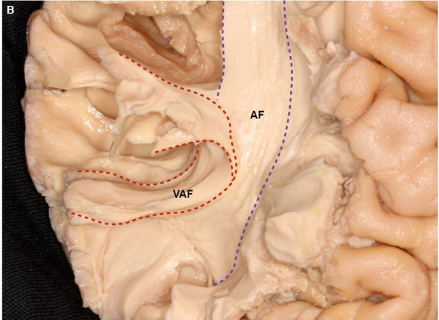
Parietal aslant tract

View Details
Status
Done

General

  • Aka: Vertical Arcuate Fasciculus
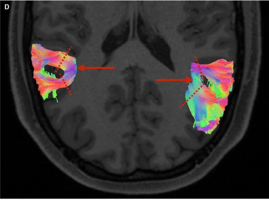
  • No lateralization (equal in each hemisphere)
  • A distinct anterior-posterior bifurcation pattern
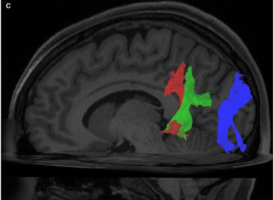
Two segments

  • Ventral VAF (vVAF)
    • Preferential SmG-MTG connectivity may therefore serve as a conduit for mapping sounds or read words to their meanings, the latter function potentially being subserved by MTG
  • Dorsal VAF (dVAF)
    • Rightward-preferential AG-MTG/ITG connectivity, may thus subserve roles in visuospatially locating objects in a 3D space.
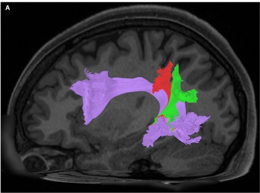
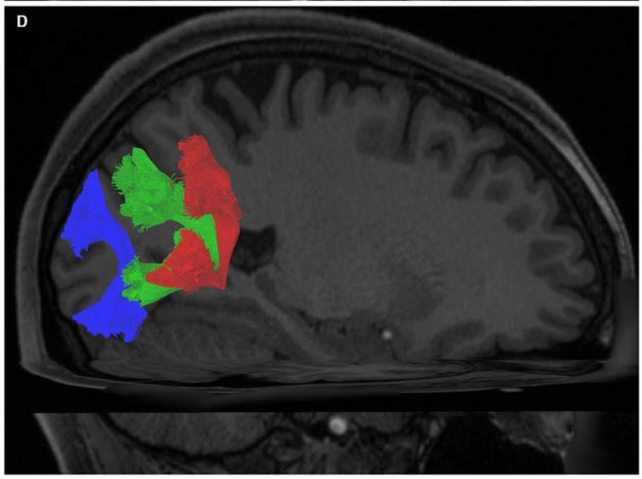
Course

  • Travel vertically and dorsal to the Sylvian fissure to their parietal terminations

Connectivity

  • Between superior marginal gyrus ←→ Middle temporal gyrus

Images

A sagittal view of the left hemisphere of a single subject. This image demonstrates the relationship of the vVAF (red) and dVAF (green) to the underlying AF (purple).
Superior axial view of posterior cerebral hemispheres, demonstrating bilateral VAFs. The red arrows indicate bifurcation of the left and right bundles, respectively. The dashed red lines represent the trajectory at which the anterior and posterior bundles were subsequently selected.
Sagittal view of the left hemisphere of a single subject. Visible are the VAF and VOF. The VAF has been segregated manually, and separate colors have been assigned to to the vVAF (red) and dVAF (green). The VOF has been colored blue. Apparent is the anteroposterior spatial separation between the entire VAF and the VOF. 

A magnified view of a dissected VAF, with its demarcation from the AF proper marked out. The VAF is bounded by red-dashed lines, and the AF-proper is bounded by purple-dashed lines.
Sagittal view of the right hemisphere of a single subject. Visible are the VAF and VOF. The VAF has been segregated manually, and separate colors have been assigned to the vVAF (red) and dVAF (green). The VOF has been colored blue. Apparent is the anteroposterior spatial separation between the entire VAF and the VOF.