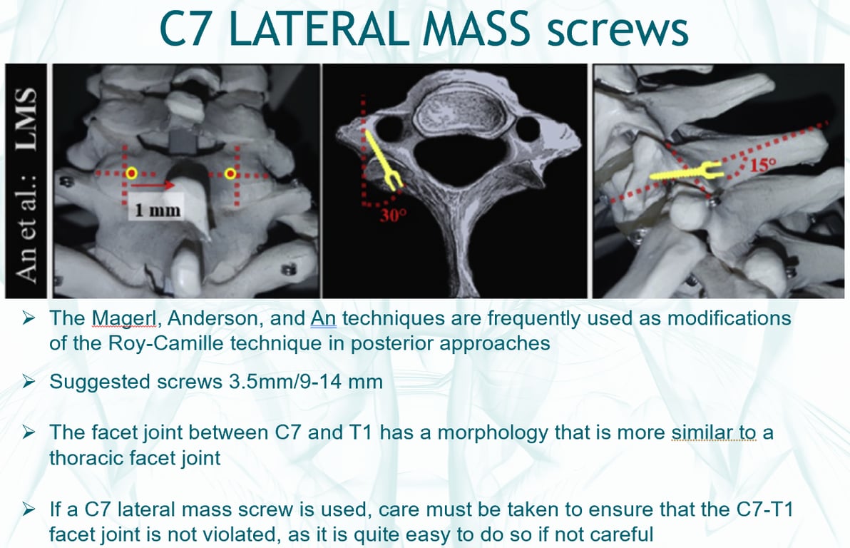
Lateral mass screws
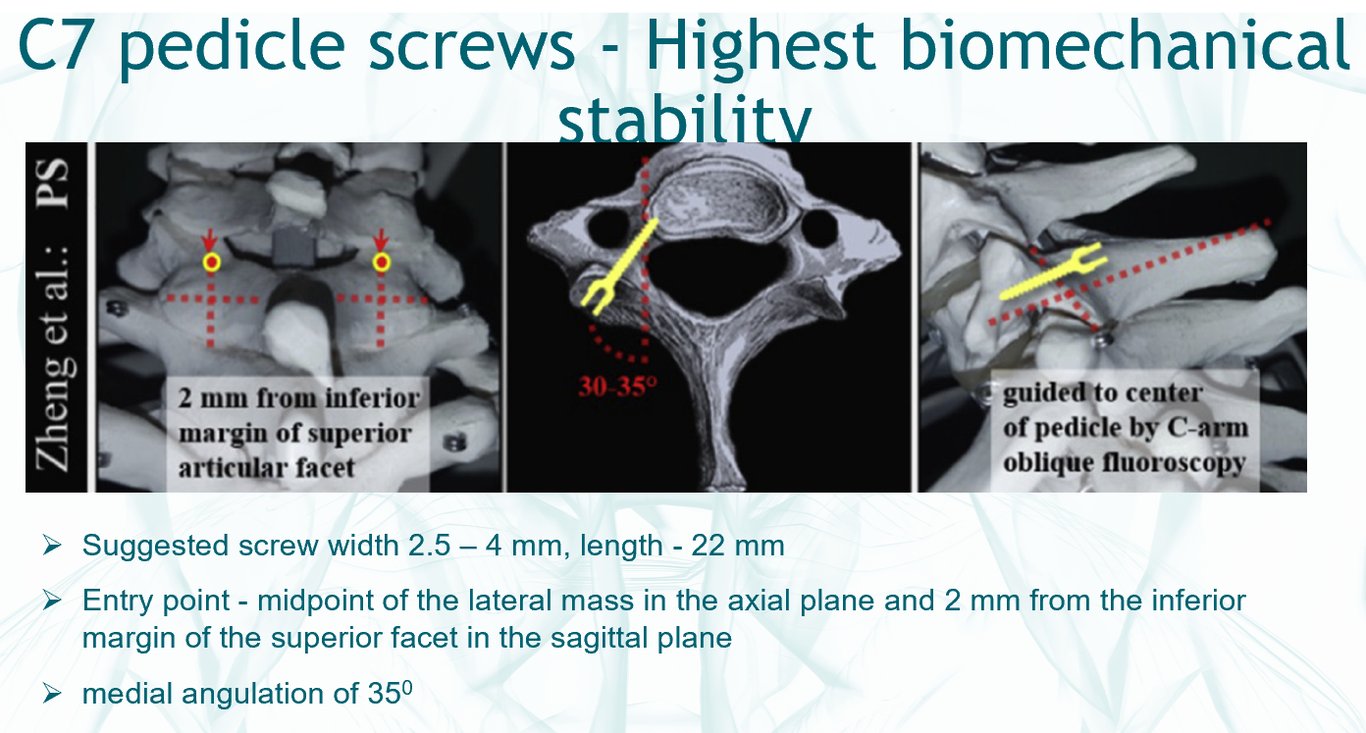
Pedicele screw
- The pedicles in the lower cervical spine (C6 and C7) may have pedicles of sufficient calibre to present a useful surgical target, the midcervical spine less so.
- presence of the vertebral artery in the foramen transversarium from C6 upwards is a complication
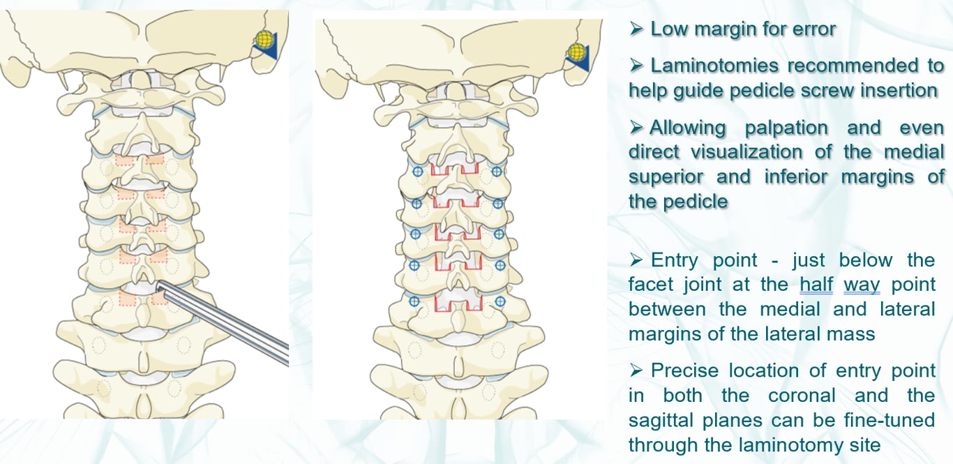
- Entry point
- Monopolar and visualize the C6/7 facetal joint
- Entry point mid point of the facet joint and just inferior to the C6/7 facet joint
- Trajectory:
- Cranial caudal angle: parallel to end plate
- medio-lateral angle:
- 90 degree to the lamina
- medial angulation of 35 deg
- AS: the direction of the C7 Pedicle (or any cervical pedicle screw) is parallell to the contralateral endplate.
- Joaquim 2020
Technique | Entry point | Lateral angulation | Sagittal angulation |
Free hand C7 pedicle screw technique (Riew technique) | At the junction of C6–7 facet joints, just below the inferior facet articular process of C6 and slightly lateral (about 2 to 4 mm) | 45° from the midline (angulation assessed using preoperative CT scan evaluation) | 90° with the superior facet joint of C7 |
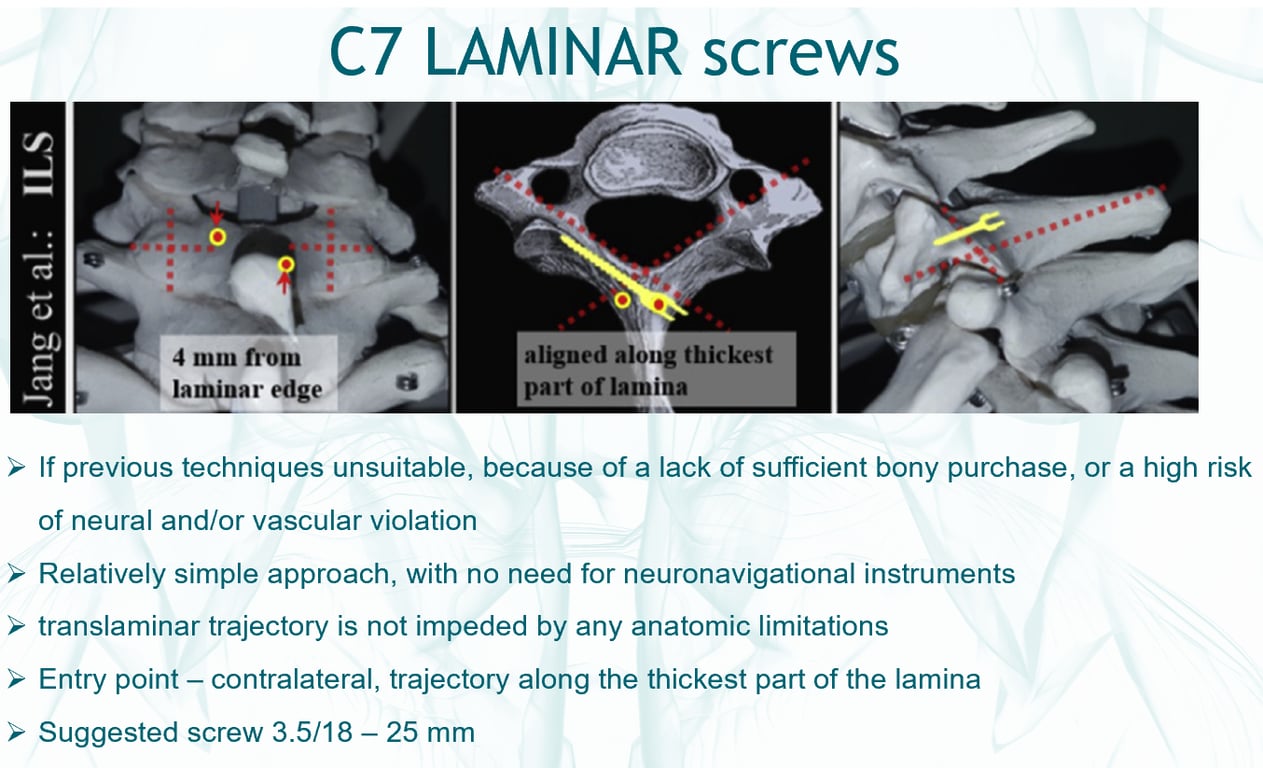
Laminar screw
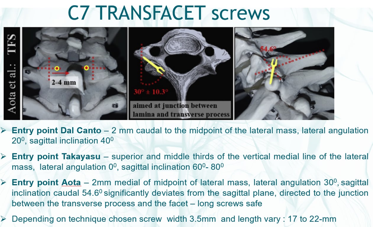
Transfacetal screw
Posterior cervical wiring
- Complication
- Injury of the vertebral artery
- Esp in an extremely small lateral mass, often at C3, making screw placement difficult or an aberrant vertebral artery course, as indicated by the foramina transversaria.
- If the surgeon has specific concerns about the course of the vertebral artery, CT angiography of the neck vasculature is indicated
- Injury of the nerve root
- Violation of the facet joints
- Pedicle screw
- Posterior cervical wiring and transfacet screws
- Salvage techniques where other methods are not possible or have failed.
 Made with Bullet