General
- Intervertebral Micro Access Surgery (iMAS), a novel hybrid surgical technique that integrates elements of both traditional open surgery and minimally invasive surgery (MIS) for performing transforaminal lumbar interbody fusion (TLIF).
- Hybrid Nature: iMAS utilizes a traditional midline skin incision but employs a paramedian approach to access the facet complexes, minimizing muscle disruption similar to standard MIS techniques.
- The Interpedicular Space: The paper defines this "work space" as the 3-dimensional area bounded by the superior pedicle, inferior pedicle, and the midline. To enhance precision, the authors identify seven unique 3-dimensional zones within and around this space to guide decompression and instrument placement.
Anatomy
- Zones 1 to 3 represent the preforaminal and foraminal zones.
- Zone 4 is the extraforaminal inferior zone. It is located at the lateral margin of the lateral facet, within the inferior interspace between the 2 pedicles and is significant for extreme lateral disc herniations, osteophytes, laterally oriented synovial cysts, and laterally migrated intervertebral implants.
- Zones 5 and 6 represent the working space for the facetectomy, dis- cectomy, and subsequent interbody fusion.
- Zone 7 is the central zone that may be significant ventrally for central disc herniations and/or posteriorly, if there is significant ligamentum flavum hypertrophy.
Technique
- GA
- Anesthesia induction must avoid long-acting muscle relaxants to allow for mandatory real-time electromyography (EMG) monitoring.
- Positioning
- AP and lateral fluoroscopy are used to mark the pedicle entry points and establish regional alignment.
- Incision and Fascial Release
- A midline skin incision (typically 2–3 cm) is made, followed by an epifascial dissection to allow the skin to be maneuvered over the working channel.
- Bilateral fascial incisions are made 1–3 cm lateral to the midline to create an access corridor while sparing the medial multifidus muscle attachments.
- Pedicle Screw Insertion
- Pedicles are cannulated using AP fluoroscopy or neuronavigation, typically starting at the lowermost lateral quadrant of the pedicle.
- The use of a headless screw system is preferred to keep the workspace clear for the later interbody fusion; these screws serve as a "grid map" for the 3D architecture of the interpedicular space.
- Facetectomy for Interbody
- Under microscopic visualization, a box-shaped facetectomy is performed to provide access to the disc space.
- Fluoroscopy combined with visual cues from the pedicles allows marking of the rostral and caudal extent of the box facetectomy that will allow access to the disc space.
- The caudal screw provides an anatomic reference for the medial working space.
- The medial extent of the working channel will be approximately 0.5 to 1.0 cm medial to the caudal screw, whereas the rostral extent varies depending on the degree of disc space collapse.
- Care should be taken to leave a rim of cortical bone medially to protect the lateral dura while maintaining the rostral/lateral bone to protect the exiting nerve root
- Transforaminal Interbody Implant Placement
- The disc is removed and end plates are prepared using specialized curettes and shavers under fluoroscopic guidance.
- A banana-shaped cage or steerable interbody device is inserted; nerve monitoring is maintained throughout this stage by passing electrical current through the instruments to detect proximity to the nerve root.
- Direct Decompression
- This step utilizes an "out-in" approach, starting with a lumbar keyhole opening located caudal and medial to the rostral pedicle.
- The surgeon moves caudally and medially to remove the lateral lamina and ligamentum flavum, allowing for direct visualization and decompression of the thecal sac and nerve roots.
- Bone to be removed as the decompression moves from lateral rostral to caudal medial.
- Anteroposterior illustration of further rostral decompression of foraminal exiting nerve root, (red circle) illustrating exiting nerve root decompressed.
- Rod/Screw Cap Assembly and Compression
- If headless screws were used, polyaxial heads are now affixed to them.
- Rods are slid beneath the fascia and seated into the screw heads using fluoroscopy, followed by the placement of locking caps and the application of compression.
- Decortication
- To prepare for posterior fusion, the surgeon clears soft tissue from the contralateral (non-decompressed) side.
- A drill is used to aggressively decorticate the contralateral facet joint and the lamina where it meets the spinous process.
- Onlay Fusion
- Autograft and/or allograft is packed medial to the screw/rod construct on the decorticated surfaces to facilitate a robust posterior arthrodesis.
- Closure
- Both fascial layers are closed to the medial spinous process to eliminate dead space.
- The skin is closed with dermal sutures and surgical adhesive (Dermabond), resulting in a small postoperative footprint.
Surgical tools
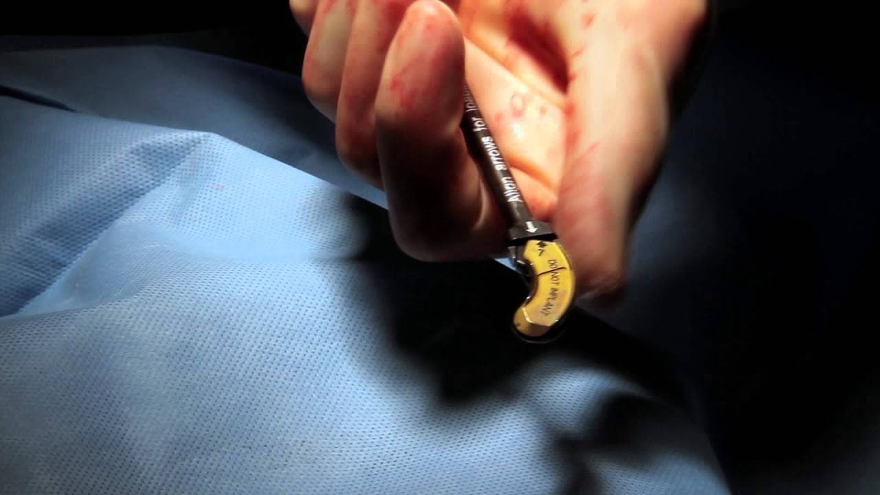
- Headless Pedicle Screw System:
- This preserves a wider working corridor for the discectomy and interbody fusion, with the polyaxial screw heads only being attached just before rod placement.
- Grid Mapping:
- Previously placed screws serve as visual anatomic cues, providing a "grid map" that helps the surgeon maintain orientation within the small working corridor.
- Nerve Monitoring:
- To avoid nerve injury during instrument passage, the authors use real-time electromyography or a method of passing electrical current through instruments to trigger visible motor activity in the patient's leg.
Indications
- Grade I spondylolisthesis
- Synovial cysts with instability
- Recurrent disc herniations
Advantages
- Minimal tissue disruption, which may lead to more rapid recovery, reduced blood loss, and shorter hospitalisations compared to traditional open surgery.
Disadvantages
- Steep learning curve
- Significant radiation exposure from the heavy reliance on fluoroscopy.
- Body mass index (BMI) over 35 present unique technical challenges, often leading to longer operative times.