- Optical aids (e.g. loupes, OMs, surgical headlamps, fiberoptic handpiece lights) can improve resolution by many orders of magnitude.
- For example, a common operative microscope can raise the resolving limit from 0.2 to 0.006 mm (6 μm), a dramatic improvement.
- Stereopsis, or 3D perception, is critical to achieving precision surgery, and is an advantage over 2D endoscopes.
- Several factors are important for increasing resolution without compromising ergonomics, eyestrain, head/neck fatigue
- Allows stereoscopic vision
- The separation of the reflected light into two beams within a microscope is what produces the stereoscopic effect that allows the clinician to see depth of field.
- The optical unit consists of:
- Eyepieces
- Magnify the interim image generated in the binocular tubes. Eyepiece selection not only determines the magnification, but also the size of the field of view.
- Binoculars
- The precise adjustment of the interpupillary distance (by adjusting the distance between binocular tubes holding the eyepieces) is the basic pre-requisite for the stereoscopic view of the operation area. Longer the focal length of binoculars, the greater the magnification and narrower the field of view. Many microscopes now include a beam splitter and a second set of teaching binoculars (non-stereoscopic as they split light from a single objective).
- Magnification changer (binocular objectives)
- One cylinder, into which two Galilean telescope systems with various magnification factors are built.
- The combination of the magnification changer with varying objective lenses and eyepiece yields an increasing magnification line when the control is adjusted.
- Objective lens
- Its focal length determines the working distance between the lens and the surgical field.
- Magnification of approximately 4-40 × with an excellent illumination of the working area.
- Focal Length:
- The distance between the eye/lens and the patient
- As the focal length decreases, the eyes must converge more closely around an axis to reduce the depth of field. → eye strain
- Working distance
- The distance from the microscope objective lens to the point of focus of the optical system.
- This value is fixed and is dependent on the chosen focal length of the objective lens
- Resolving power:
- Ability to make clear and distinguishable two separate entities.
- The resolving power of the unaided human eye is only 0.2 mm.
- Most people who view two points closer than 0.2 mm will see only 1 point. Moving closer to an object increases resolution up to a point, but objects closer than 10-12 cm go out of focus.
- Depth of Field:
- The far-near range within which objects remains in focus without having to adjust working distance.
- As magnification increases, depth of field decreases hence focusing on multiple objects at different depths with high magnification will require more changes in microscope position.
- Also, the smaller the field of view, the shallower the depth of field
- Width of Field:
- The width and height of the area visible through a lens system, which decreases with increasing magnification
- Lighting unit
- As optimal illumination is necessary with high magnifications.
- The light beams fall parallel onto the retinas of the observer so that no eye convergence is necessary and the demand on the lateral rectus muscles is minimal.
- Surgical microscope uses coaxial fiber-optic illumination producing an adjustable, bright, uniformly illuminated, shadow-free, circular spot of light that is parallel to the optical viewing axis.
- By increasing light levels, one can increase Resolving power: (the ability to distinguish two objects close to each other as separate and distinct).
- Light intensity is determined by the inverse square law, which states that the amount of light received from a source is inversely proportional to the square of the distance
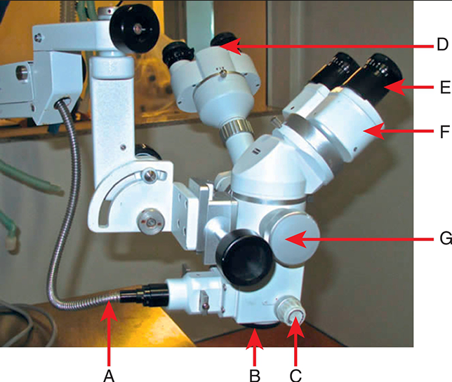
A = light fiber;
B = objective lens;
C = magnification changer;
D = teaching binoculars;
E = eyepiece;
F = main binoculars;
G = beam splitter
 Made with Bullet