Definition
- CSF-containing cyst covered by arachnoidal membranes which are continuous with the normal surrounding arachnoid.
Numbers
- prevalence in adults is approximately 1.4%
- female preponderance,
- prevalence in children is 2.6%
Aetiology
- Primary cysts
- Arise from the splitting of the arachnoid membranes in utero, resulting in the development of anomalous collections of CSF
- Secondary cysts
- Are less common
- Often appearing after trauma, surgery, infection, or intracranial haemorrhage
Mechanism
- Normal
- At 15 wks, the rhombic roof ruptures, CSF pulses through the meninx primitive (loose connective tissue precursor to pia and arachnoid lining surface of dura) forming the subarachnoid space as the pia mater and arachnoid separate incompletely, resulting in the cobweb-like appearance of the arachnoid.
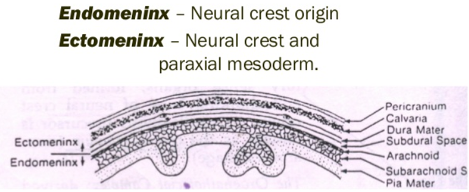
- Abnormal
- When the normal separation of the arachnoid and pia is abnormally enclosed, loculated chambers form and develop into a cystic mass (arachnoid cyst).
- Abnormal splitting and duplication of the endomeninx, which normally forms a loose extracellular substance in the future CSF-filled subarachnoid space.
- The Endo and ectomenix are formed from the primitive meninx
- The primitive meninx is the origin of intracranial lipomas
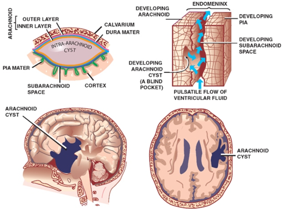
- Why Arachnoid cyst expand
- Slit-valve or ball-valve communication may exist between the subarachnoid space and the cyst, allowing entry but not egress of CSF during normal CSF pulsation or Valsalva manoeuvres and the local geometry of the split membrane may provide the pressure gradient to drive fluid into the cyst.
- Slit valves have been directly observed with endoscopy and currently are the most direct explanation for why the cysts expand.
- Located in an arachnoid rich cisterns
Clinical features
- Depend on the
- size of the cyst
- Small cysts are often incidental
- Large cyst are symptomatic
- Mass effect
- Raised intracranial pressure
- Location
- Cysts in the chiasmatic region can produce
- Visual disturbances
- Endocrine disorders
- Compromise of the hypothalamus-pituitary
- 3rd/4th ventricle compression
- HCP
- can cause bone remodelling
- in young children may produce
- Macrocrania
- Cranial asymmetry
- associated with delayed psychomotor development
- Subdural hematoma
- Intracystic haemorrhage
- Acute cyst expansion
- Subdural hygroma
Radiology
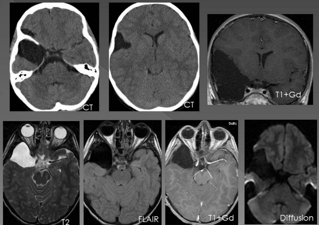
- Cyst has the same signal intensity as CSF at all MRI sequences
- Does not enhance with contrast administration.
- Produces a discrete mass effect on frontal lobe and the right temporal lobe is hypoplastic.
- Right middle cerebral artery is slightly displaced by the cyst
- MRI cine might show communication with subarachnoid space
Management
- General
- The actual rate of cyst-related intracranial haemorrhage after contact sports-related head injury is essentially unknown, so can use any options to tx
- Conservative
- Surgery
- Arguments for surgery
- Theoretical risk of spontaneous or traumatic intracranial bleeding)
- Craniotomy and microscopic
- Endoscopic fenestration
- Needle aspiration
- Cystoperitoneal shunting
 Made with Bullet