Number
- 50-65% of all arachnoid cyst
Galassi classification
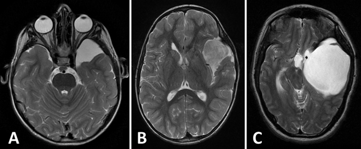
- Type I:
- small size, located in the anterior temporal lobe.
- No mass effect
- Free communication with Subarachnoid space
- Tx with microsurgical fenestration
- Type II:
- medium-size, located in the anterior and middle temporal fossa. Temporal lobe displaced.
- Slow communication with Subarachnoid space
- Tx with endoscopic fenestration
- Type III:
- constitutes a large oval or round cyst that fills the entire temporal fossa. Large mass effect.
- Little communication with Subarachnoid space
- Tx with endoscopic fenestration
Tx
- Surgical: no difference in all 3
- Endoscopic,
- endoscopic fenestration is the preferred primary surgical modality.
- Microsurgical,
- Only be considered when symptoms are unchanged after endoscopic treatment
- Shunting
- Only be considered when symptoms are unchanged after endoscopic treatment
- ICP monitoring useful for T1 and T2 but not T3
- To may help to rule out operative treatment in Type I, confirm raised ICP in Type III (but inconsistent in type II).
 Made with Bullet