General information
- A failure of commissuration occurring ≈ 2 weeks after conception --> expansion of the third ventricle and separation of the lateral ventricles (which develop dilated occipital horns and atria, and concave medial borders).
- The corpus callosum (CC) forms from rostrum (genu) to splenium, ∴ in agenesis there may be an anterior portion with absence of the posterior segment (the converse occurs infrequently).
- Absence of the anterior CC with presence of some posterior CC is indicative of some form of holoprosencephaly.
- May be an incidental finding, and by itself may have no clinical significance.
Numbers
- Incidence 1 in 2,000–3,000 neuroradiological examinations.
Associated neuropathologic findings
- Porencephaly
- Microgyria
- Interhemispheric lipomas and lipomas of the corpus callosum (p.276)
- Arhinencephaly
- Optic atrophy
- Colobomas
- Hypoplasia of the limbic system
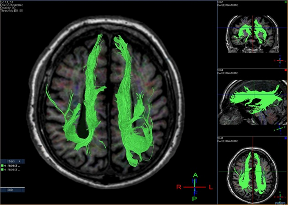
- Bundles of Probst: aborted beginnings of corpus callosum, bulge into lateral ventricles
- loss of horizontal orientation of cingulate gyrus
- Schizencephaly (p.304)
- anterior and hippocampal commissures may be totally or partially absent44
- hydrocephalus
- cysts in the region of the corpus callosum
- spina bifida with or without myelomeningocele
- absence of the septum pellucidum
- Aicardi syndrome
Possible presentation
- Hydrocephalus
- Microcephaly
- Seizures (rare)
- Precocious puberty
- Disconnection syndrome: more likely with acquired CC defect than with congenital
Anomalies of the Corpus Callosum
- Normal corpus callosum development
- Rostral to caudal sequence
- Genu → Body → Splenium
- Exception: rostrum which develop last.
- Normally, the corpus develops between the 8th and 20th weeks of gestation, at the same time as the rest of the cerebrum and cerebellum.
- Abnormal
- Hypogenesis of the corpus callosum due to developmental arrest produces an intact Genu and body with an absent splenium and rostrum
- Any other pattern than the one stated above means that the corpus callosum abnormality is due to a destructive process which has occurred after the normal formation of the corpus callosum.
- Corpus demonstrates an intact splenium in the absence of a genu or body.
- Because the corpus develops at the same time as the cerebrum and cerebellum, callosal anomalies are often associated with other brain anomalies
- Dandy-Walker malformation
- Disorders of neuronal migration and organization
- Encephaloceles
- Most common type of commissural agenesis
- Associated with absence of the hippocampal commissure
- MRI
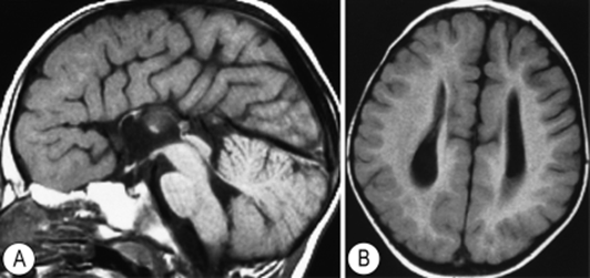
- Mid-sagittal MR images are diagnostic
- Features
- Everted cingulate gyrus
- Longitudinal Probst bundles containing non-crossing callosal axons
- Probst bundles or longitudinal callosal fascicles denote white matter fibres normally destined to cross the corpus callosum that instead parallel the interhemispheric fissure
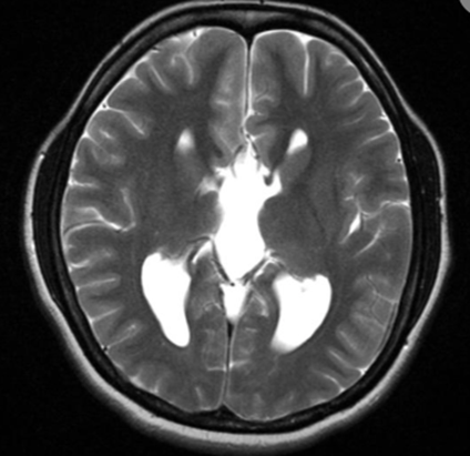
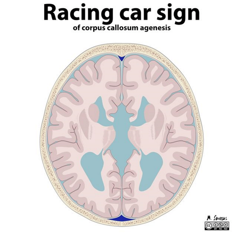
- lateral ventricles are shifted laterally and closed medially by rolled up white matter lamina (which should be forming the leaf of the septum pellucidum).
- The inner walls of the lateral ventricles are concave medially as a result of encroachment of the Probst bundles on the ventricular lumen.
- Roof of the third ventricle bulges upward.
- The frontal horns of the lateral ventricles might be underdeveloped, whereas the dilated temporal horns invaginate into the core of the parahippocampal gyri because of decreased white matter.
- The lateral ventricles run parallel to each other, with marked dilation of the trigone and occipital horns (colpocephaly).
- Associated with
- Chiari malformation
- Dandy-Walker malformation
- Neuronal migration anomalies
- Periventricular or subcortical heterotopia
- Midline facial anomalies (facial cleft, encephalocele)
- Aicardi's syndrome
- An X-linked disorder
- Sporadic condition
- Triad of
- Total or partial agenesis of the corpus callosum
- Infantile spasms
- Chorioretinal lacunae
- Well-defined, punched-out lesions in the pigmented layer of the retina, most commonly found around the optic disc.
- They represent areas where both the retina and the underlying choroid are absent or markedly atrophic, creating distinctive patches that appear darker upon examination
- Abnormal electroen-cephalogram use to support diagnosis
- Intact genu
- Partially or completely formed body
- Small or absent splenium and rostrum
- Will lose the above sequence
- Eg: a small or absent genu or body but an intact splenium and rostrum,
- Clinical features
- Isolated anomalies of the corpus callosum are usually asymptomatic.
- Symptoms, when present, are often related to associated brain anomalies.
- The most common associated symptoms are seizures and mental retardation.
2 types of Developmental abnormalities:
Callosal anomaly associated with holoprosencephaly
Developmental arrest of the corpus may result in its partial or complete absence.
Complete agenesis of corpus callosum
 Made with Bullet