General features
- 1.5% of encephaloceles
- The only group that does not produce a visible soft tissue mass.
- Present as CSF leak or recurrent meningitis.
- Associated with other craniofacial deformities, including: cleft lip, bifid nose, optic-nerve dysplasia, coloboma and microphthalmia, hypothalamic-pituitary dysfunction.
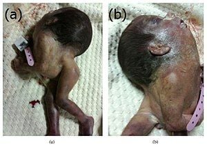
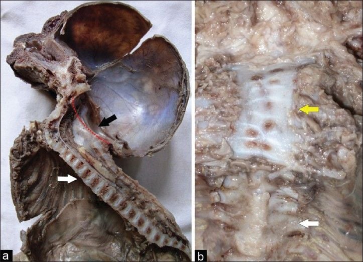
- Iniencephaly
- Caused by improper closure of the neural tube
- Characterized by defects around the foramen magnum, rachischisis and retrocollis (due to fused vertebrae).
- Most are stillborn, some survive up to age 17
Transethmoidal
- Protrudes into nasal cavity through defect in cribriform plate
Spheno-ethmoidal
- Protrudes into posterior nasal cavity
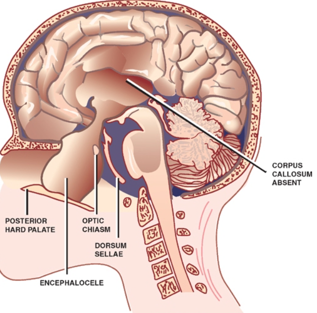
Transsphenoidal
- Protrudes into sphenoid sinus or nasopharynx through patent craniopharyngeal canal (foramen cecum)
- Nasopharyngeal (either spheno-ethmoidal or transphenoidal)
- Rare.
- Diagnosed during an evaluation for persistent nasal stuffness or excessive “mouth breathing.”
- Examination
- Nasopharyngeal masses that increase in size with a Valsalva maneuver.
- Associated intracranial anomalies
- Callosal agenesis (common)
- Clinical features
- Endocrine and visual dysfunction
- Due to tethering of the hypothalamus and optic chiasm as they extend into the sac
Fronto-sphenoidal or spheno-orbital
- Protrudes into orbit through superior orbital fissure
 Made with Bullet