General
- AKA arhinencephaly.
Definition
- A group of disorder due to failure of differentiation and cleavage of the prosencephalon.
Numbers
- most common developmental defect of the forebrain
- A live birth prevalence of approximately 1 in 10,000
Genetics
- 80% are associated with trisomy (primarily trisomy 13, and to a lesser extent trisomy 18).
Mechanism
- Normal
- 32 day: horizontal cleavage
- Germinal matrix begins to cleave into superior and inferior portions.
- Superior portion form the telencephalon (the caudate, putamen, and cerebral hemispheres)
- Inferior portion will give rise to neurons that form the diencephalon (the thalamus, hypothalamus, and globus pallidus).
- Disease
- prosencephalon cannot separate in to the telencephalon and the diencephalon
- 35 day: vertical cleavage
- Evagination and separation of the cerebral hemispheres
- This division takes place as a result of induction by bone morphogenetic protein from the midline roof plate.
- 32-34 day lamina terminalis begins to differentiate into the inter-hemispheric cerebral commissures (anterior commissure, corpus callosum and fornix commissure)
- Disease: MAIN CAUSE OF HOLOPROSENCEPHALY
- Germinal matrix and the lamina terminalis cannot separate into two hemispheres.
- Non-cleavage of midline ventral forebrain at 33 days --> failure of hemispheric separation
Aetiology
- Genetic
- Trisomy 13 (Patau syndrome)
- SHH mutation
- SHH is a protein that encodes a morphogen which mediates notochordal-ventral neural tube and development of craniofacial structures (facial deformities are also seen in association with the HPE spectrum).
- Environmental
- maternal diabetes
- Exposure to teratogens such as alcohol.
Clinical features
- Abnormal synaptogenesis is early causing
- Early development of the median cyclopean eye
- Formation of epileptogenic circuitry
- Severe infantile epilepsy
- Ohtahara syndrome
- Epileptic (infantile) spasms or west syndrome with hyperarrhythmic electroencephalogram
Pathology
- The olfactory bulbs are usually small and the cingulate gyrus remains fused.
- Median faciocerebral dysplasia is common,
- Degree of severity = extent of the cleavage failure
- Five facies of severe holoprosencephaly
Type of face | Facial features | Cranium and brain findings |
Cyclopia | Single eye or partially divided eye in a single orbit; arhinia with proboscis | Microcephaly; alobar holoprosencephaly |
Ethmocephaly | Extreme orbital hypotelorism; separate orbits; arhinia with proboscis | Microcephaly; alobar holoprosencephaly |
Cebocephaly | Orbital hypotelorism; proboscis-like nose; no median cleft lip | Microcephaly; usually has alobar holoprosencephaly |
With median cleft lip | Orbital hypotelorism; flat nose | Microcephaly; sometimes has trigonocephaly; usually has alobar holoprosencephaly |
With median philtrum-premaxilla anlage | Orbital hypotelorism; bilateral lateral cleft lip with median process representing philtrum-premaxillary anlage; flat nose | Microcephaly; sometimes has trigonocephaly; semilobar or lobar holoprosencephaly |
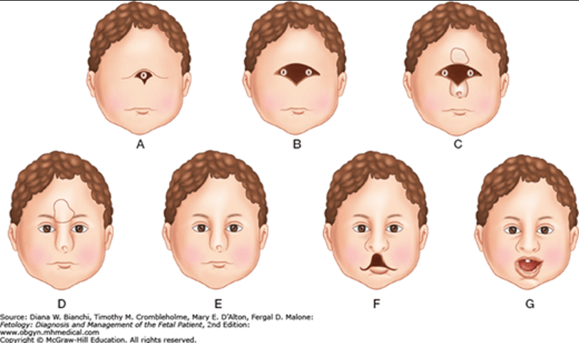
C shows cyclopia with a proboscis.
D shows ethmocephaly with a proboscis.
E demonstrates cebocephaly with a single nostril.
F shows mild holoprosencephaly with a midline cleft lip.
G is the mildest form of holoprosencephaly with a single central incisor
Subtypes: provides the degree of cleavage failure
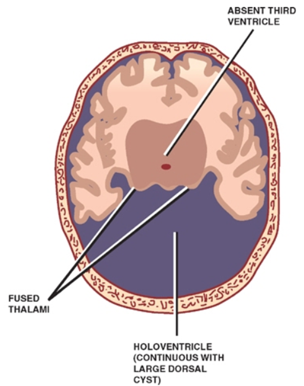
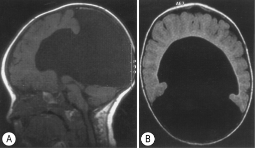
Alobar holoprosencephaly (0205198465)
- Most severe form
- Most common
- Complete failure in forebrain separation resulting in single holospheric cerebrum
- Holosphere
- remains undivided as a single flattened mass of brain surrounding a midline holoventricle that is large and shaped like an inverted “U” or crescent.
- displaced in the most cephalad part of the intracranial cavity.
- Fused basal ganglia, thalamus
- located in the floor of the holoventricle.
- No interhemispheric fissure, falx cerebri, or corpus callosum, Gyri recti, no 3rd ventricle or sylvian fissure
- A holoventricle is contiguous with a large dorsal cyst, leaving only a small rim of brain anteriorly.
- Clinical features
- stillbirth or a very short lifespan
- Associated anomalies
- Severe midline facial deformities
- premaxillary agenesis, cleft lip/palate
- Hypotelorism, which in its most severe form is manifest by cyclopia.
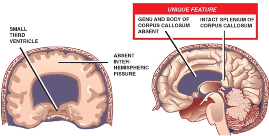
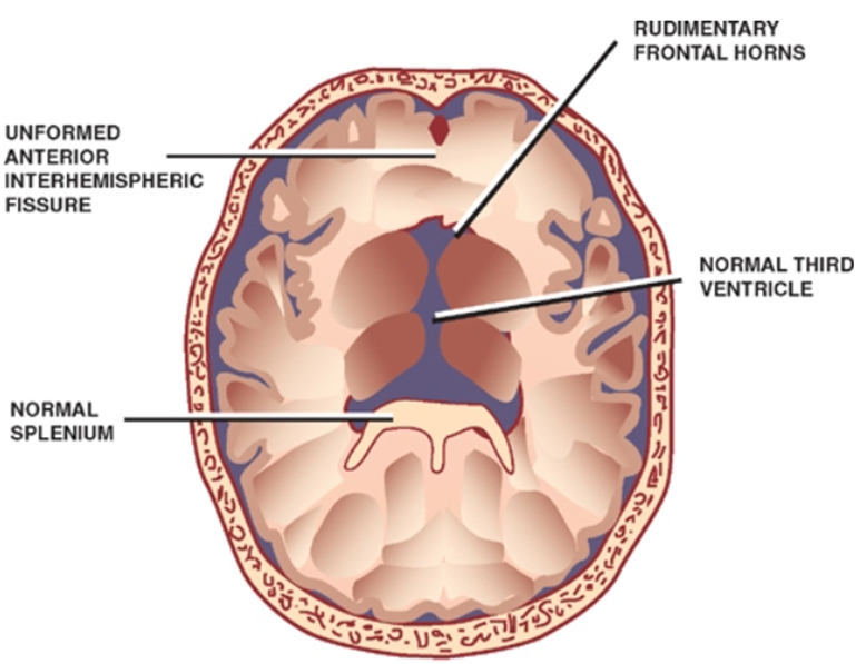
Semilobar holoprosencephaly
- Less severe
- Frontal and parietal lobes appear fused but posterior interhemispheric fissure present.
- At least a partial separation of the thalami, and thus a small third ventricle
- A partially formed or absent interhemispheric fissure and falx cerebri.
- Intact splenium but a small or absent genu and body.
- In contrast to the abnormal development sq of the corpus callosum
Lobar holoprosencephaly
- Less severe form
- only rostral most areas of cerebral hemispheres show fusion.
- fully formed third ventricle
- intact corpus callosum.
- Septum pellucidum is absent
- Same for all types of holoprosencephaly
- Frontal lobes are typically hypoplastic.
Other types
- Syntelencephaly (middle hemisphere variant)
- hemispheres separated rostrally and caudally
- Posterior frontal lobe/parietal lobe still fused.
- Arrhinencephaly
- Absent
- olfactory bulbs
- olfactory tracts
- gyri recti.
Outcome
- Survival beyond infancy is uncommon;
- most survivors are severely retarded, and a minority are able to function in society.
- Some develop shunt-dependent hydrocephalus.
- The risk of holoprosencephaly is increased in subsequent pregnancies of the same couple
 Made with Bullet