Locations
- Deep interhemispheric fissure (40-50%)
- Quadrigeminal plate cistern (30%)
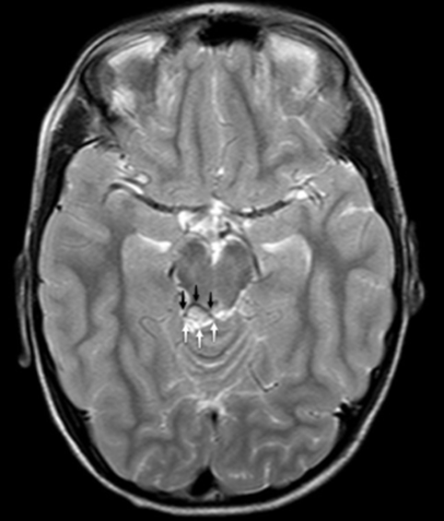
- Suprasellar/interpedicular cistern (10-20%)
- Cerebellopontine angle cistern (10%)
- Sylvian fissures (5%)
Mechanism
Normal
- In normal development, an undifferentiated mesenchyme (inner meninx primitiva) that surrounds the developing brain gives rise to the leptomeninges and the subarachnoid space.
- First to last subarachnoid space to develop
- Pre-pontomedullary cistern is the first to develop
- Cisterns around the brainstem and cerebral hemispheres
- Quadrigeminal plate
- Suprasellar system.
- Meninx primitiva
- surrounding the dorsum of the lamina terminalis is the last to become evolved.
Abnormal
- Abnormal differentiation of the undifferentiated mesenchyme (inner meninx primitive) may lead to the formation and deposition of fat in the subarachnoid space.
- Lipomas can contain blood vessels and cranial nerves, creating an obstacle to their surgical removal.
- Fq (high → low)
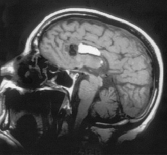
- Deep interhemispheric fissure
- Aka: lipomas of the corpus callosum
- Associated with hypogenesis or agenesis of the corpus callosum.
- There is frequently also evidence of punctate or curvilinear midline calcifications, or the presence of other anomalies, such as encephaloceles and cutaneous lipomas.
- Quadrigeminal plate cistern,
- Interpeduncular cistern,
- Cerebellopontine angle cistern,
- Sylvian cistern.
- Rarely do intra-cranial lipomas exert significant mass effect on surrounding brain structures; thus, the need for surgical intervention is rare as well.
- Because by embryologic definition lipomas occupy the subarachnoid space, blood vessels and cranial nerves course through them.
Clinical features
- Most are asymptomatic
- diagnosed incidentally.
Imaging
- CT scan
- lipoma is a well-defined, fat density mass within a cistern.
- MR appearances
- T1 hyperintensity
- T2 hyperintensity,
- Fat suppression and no enhancement.
- Chemical shift artefact seen around the hyperintensity confirms the fatty origin of the mass as opposed to haemorrhage.
- Small lipomas might not demonstrate chemical shift artefact. In such cases, fat saturation can be very helpful in differentiating this lesion from other T1 bright lesions.
- A pericallosal lipoma might also show multiple signal voids because of a combination of traversing vessels and calcification.
 Made with Bullet