General
- Malformation: a congenital morphologic anomaly of a single organ or body part due to an alteration of the primary developmental program caused by a genetic defect
- Causes
- Genetic/inherited cause
- De novo
- (ie, new in the affected child, rather than present in or transmitted by the parents) or inherited from the parents.
- Inherited mutations
- are transmitted in different patterns
- Eg
- Autosomal recessive,
- from an unaffected mother or father to her or his child),
- X linked
- From an affected mother to her son
- autosomal dominant
- from an affected mother or father to her or his child
- mitochondrially inherited diseases
- from an affected mother to her child
- Disruption: a congenital morphologic anomaly caused by the breakdown of an anatomic structure that had a normal developmental potential
- Causes
- Infection
- Haemorrhage
- Ischaemia
- Definition
- Hypoplastic:
- reduced cerebellar volume
- Dysplastic:
- Abnormal cerebellar foliation, fissuration, and architecture of the cerebellar white matter
- Eg:
- Dysplastic cerebellar gangliocytoma (Lhermitte-Duclos disease):
- Hypodysplastic:
- combination of hypoplasia and dysplasia
- Ideally best not to use dandy walker complex or variant instead use the term vermis hypoplasia or global cerebellar hypoplasia
- Disorders with vermis dysplasia
- Joubert’s syndrome
- Dandy walker malformation
- Dandy walker variant
- Isolated inferior vermian hypoplasia
- Isolated cerebellar vermis hypoplasia/atrophy
Differential diagnosis
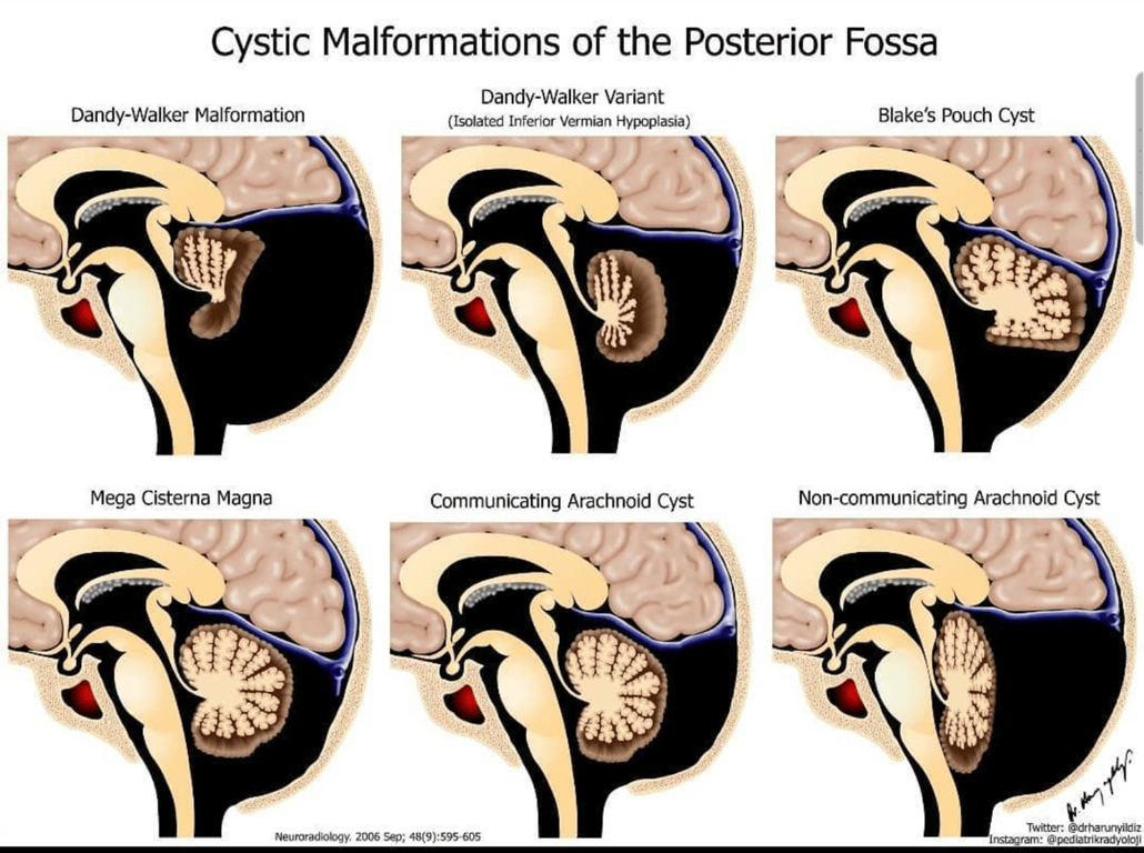
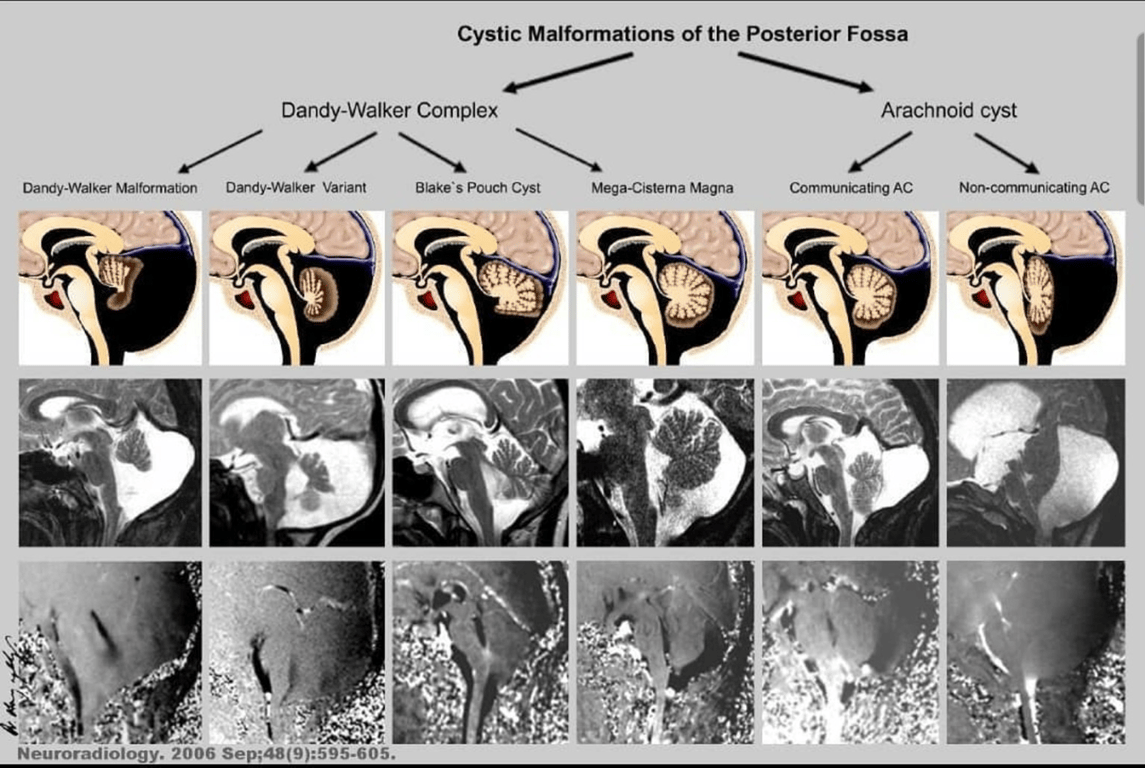
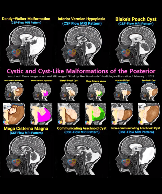
- Disorders with posterior fossa CSF (or CSF-like) collections include
- Dandy Walker malformation (DWM)
- Dandy Walker variant (DWV):
- when not all of the Dandy Walker criteria are present. E.g. vermian hypoplasia and cystic dilatation of the 4th ventricle, without enlargement of the posterior fossa
- DWM and DWV are difficult to distinguish, and may represent a continuum of developmental anomalies that are grouped together as Dandy Walker complex (DWC).
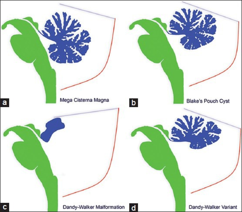
- persistent Blake’s pouch cyst (BPC):
- tetraventricular hydrocephalus, communicating 4th ventricle and posterior fossa cyst. The vermis and medial aspects of the cerebellar hemispheres may appear hypoplastic, but they usually expand after shunting (although some atrophy from the pressure may occur)
- Difference vs DWM
- do not have vermian agenesis --> Vermis is intact
- position of the choroid plexus of the 4th ventricle is displaced into the superior cyst wall in BPC, absent in Dandy Walker malformations
- retrocerebellar arachnoid cyst:
- Difference vs DWM
- do not have vermian agenesis --> Vermis is intact
- position of the choroid plexus of the 4th ventricle is normal in arachnoid cysts,
- Fourth ventriculocele
- Large posterior fossa cyst which remodels, thins and eventually erodes through the occipital bone to form an occipital encephalocoele.
- It may be classified as part of the Dandy-Walker continuum, but this is controversial.
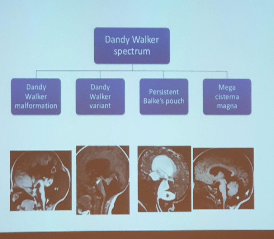
- Dandy walker complex: a spectrum of disorders made up of the following
Mild | Moderate | Severe |
mega–cisterna magna only | mild hypoplasia of vermis enlarged 4th ventricle | agenesis of vermis dilation of posterior fossa cyst and 4th ventricle |
Mega cisterna magna Blake's pouch cyst | Dandy walker variant | Dandy walker malformation |
Posterior fossa cyst comparison
Types of posterior fossa cyst
Inferior Vermis
Vermis remnant
Intrathecal contrast CT
cerebellar hemispheres
Mass effect on the cerebellar hemisphere
4th ventricle size
Position of choroid plexus
post fossa size
Occipital bone scalloping
Communication between 4th and cisterna magna
Hydrocephalus
Files & media
Image explain
Status
Hypoplasia/agenesis
cephalad rotation of the vermian remnant
Communicates with ventricles
Present Anteriorly displaced
No
enlarged
Absent
enlarged torcular-lambdoid inversion (torcular is high)
No
Yes
Yes (90%)
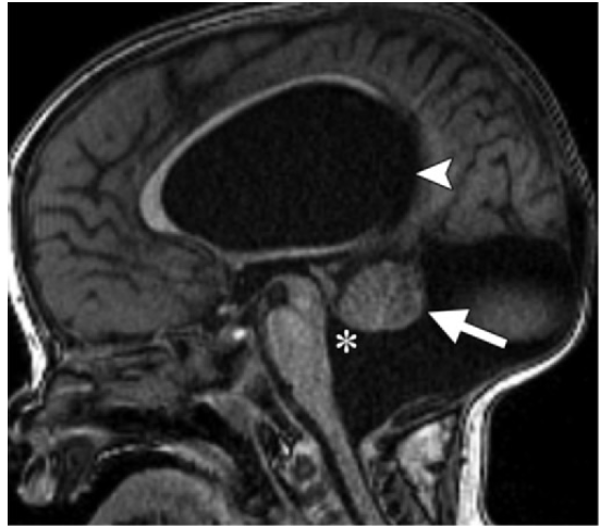
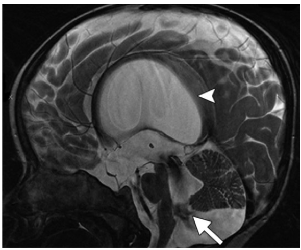
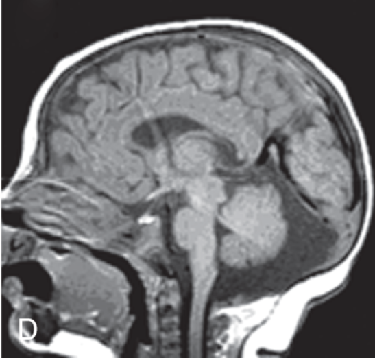
Arrow: a hypoplastic vermis in upward rotation *: cystic dilatation of the fourth ventricle Arrowhead: with posterior extension and communication with an enlarged posterior fossa, and supratentorial hydrocephalus
Done
Hypoplasia/agenesis
Communicates with ventricles
No
enlarged
absent
Normal
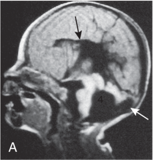
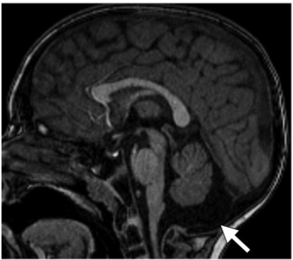
Axial T1-weighted magnetic resonance image showing Dandy-Walker variant with a fourth ventricle communicating with a retrocerebellar cyst (white arrow), absence of the inferior vermis, absence of the corpus callosum (black arrow), and lissencephaly.
Done
Normal
Normal
Communicates with ventricles
Normal
No
enlarged
displaced into the superior cyst wall
Elevation of tentorium but with a normally positioned torcula
No
No
Yes
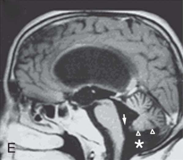
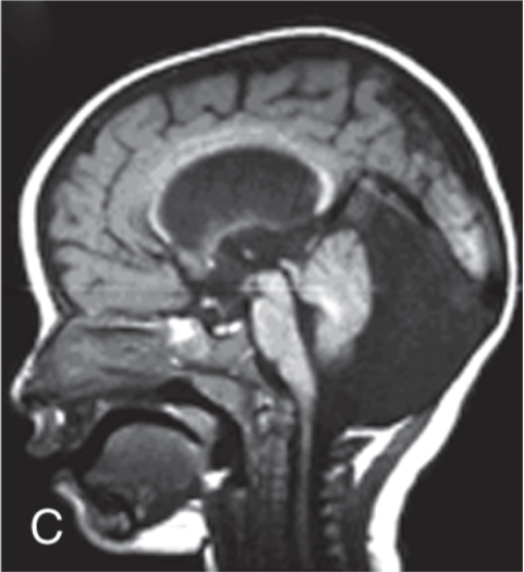
Up: • CSF collection in the fourth ventricle that is contiguous (arrow) • A collection inferior to the cerebellum (asterisk). • Small arrowheads show upward mass effect from fluid. Down: • Enlargement of 4th ventricle, which communicates with an infravermian cystic compartment (arrow) corresponding to enlargement of the Blake pouch; • a normal vermis; • and supratentorial hydrocephalus (arrowhead).
Done
Normal
Normal
May or may not communicate ventricles
Yes
enlarged
Normal position
Enlarged
Yes
Yes
anterior shift of cerebellum, which has resulted in obstruction of cerebrospinal fluid (CSF) outflow and hydrocephalus.
Done
Normal
Normal
Communicates with ventricles
Normal
No
Normal
Normal
Enlarged (some)
Possible
No
enlarged size of the posterior fossa but normal size of the cerebellum. Down: shows mega cisterna magna (arrow), a normal vermis, a normal fourth ventricle, an enlarged posterior fossa, scalloping of the occipital bone, and the absence of HCP
Done


Images
 Made with Bullet