Aka
- Isolated inferior vermian hypoplasia
Definition
- Vermian hypoplasia
- partial absence of the inferior portion of the cerebellar vermis
- Normal sized
- posterior fossa
- 4th ventricle
Presentation
- Mild functional deficits in fine motor activity and receptive language may be present.
- Inferior vermian hypoplasia in isolation has no recurrence risk;
Evaluation
Imaging
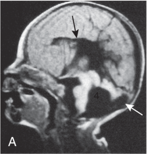
- Mid sagittal view: partial absence of the inferior vermis
- Normal size and shape
- Superior vermis
- cerebellar hemispheres,
- Fourth ventricle,
- posterior fossa
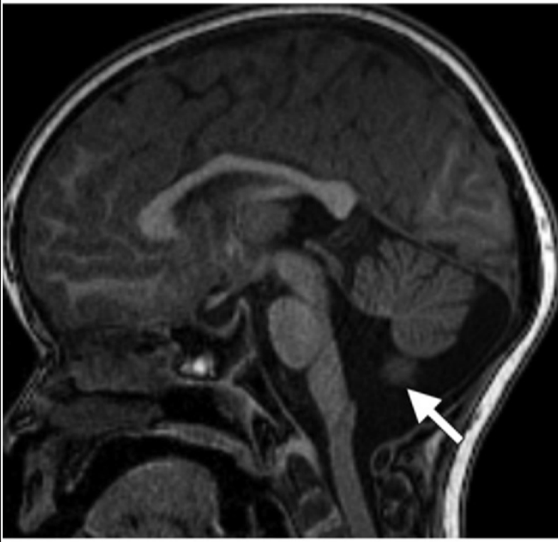
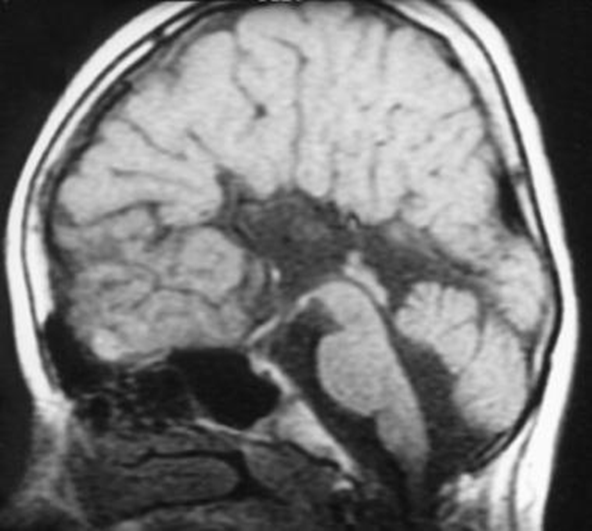
4th ventricle is slightly enlarged, but the posterior fossa typically is normal in size.
Prenatal diagnosis
- Can be done after 18-20 wks gestation using MRI as U/S is not reliable (U/S: high false positive rates 30%)
- Before 18 weeks gestation, incomplete caudal growth of the inferior vermis over the fourth ventricle may be physiologic
Outcome
- 75% of patients have a favourable outcome
 Made with Bullet