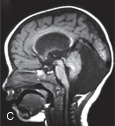
General
- See Arachnoid cyst
- Arachnoid cysts do not communicate with the fourth ventricle or the subarachnoid space.
Definition
- Duplications of the arachnoid membrane produce fluid-filled cysts known as arachnoid cysts
Numbers
- 10% of arachnoid cysts in children occur in the posterior fossa
Location
- Inferior to the vermis in a midsagittal location
- Retrocerebellar: posterior to the vermis in a midsagittal location
- Supravermian: cranial to the vermis in the tentorial hiatus
- Anterior to the cerebellar hemispheres
- Lateral to the cerebellar hemispheres
- anterior to the brainstem
Clinical presentation
- Asymptomatic → incidental finding
- Macrocephaly
- Signs of increased intracranial pressure
- By anteriorly displacing 4th ventricle and cerebellum, which can produce significant mass effect.
- Developmental delay
Evaluation
- Imaging
- MRI
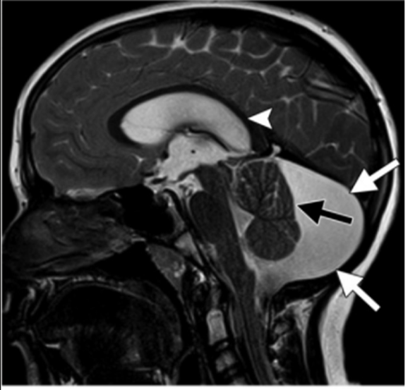
- well-circumscribed extraaxial fluid collection or cyst that is isointense relative to CSF with all sequences
- presence of proteinaceous content may lead to lack of complete signal suppression with a fluid-attenuated inversion-recovery sequence
- Cyst walls are usually too thin to be visualized at MR imaging.
- DWI
- reveals free water motion or facilitated diffusion similar to that seen in CSF
- isointense relative to CSF (white arrows), with apparent enlargement of the posterior fossa, scalloping of the occipital bone, mass effect on the dorsal aspect of a normal-appearing vermis (black arrow), a normal fourth ventricle, and supratentorial hydrocephalus (arrowhead).
Outcome
- No recurrence risk
- Favourable outcome post shunt placement
- May enlarge during infancy a
 Made with Bullet