Numbers
- most of which are below the intra-gluteal crease
- occur in as many as 4.8% of all children
- Actual problematic lesions are rare 1/2500 (spinal dysraphism)
Presentation
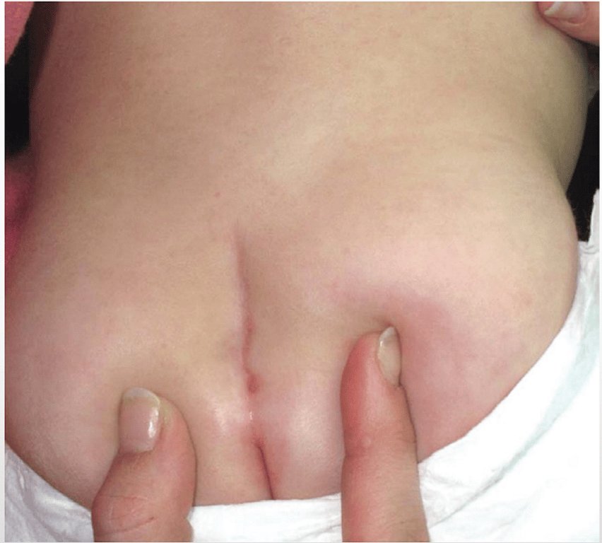
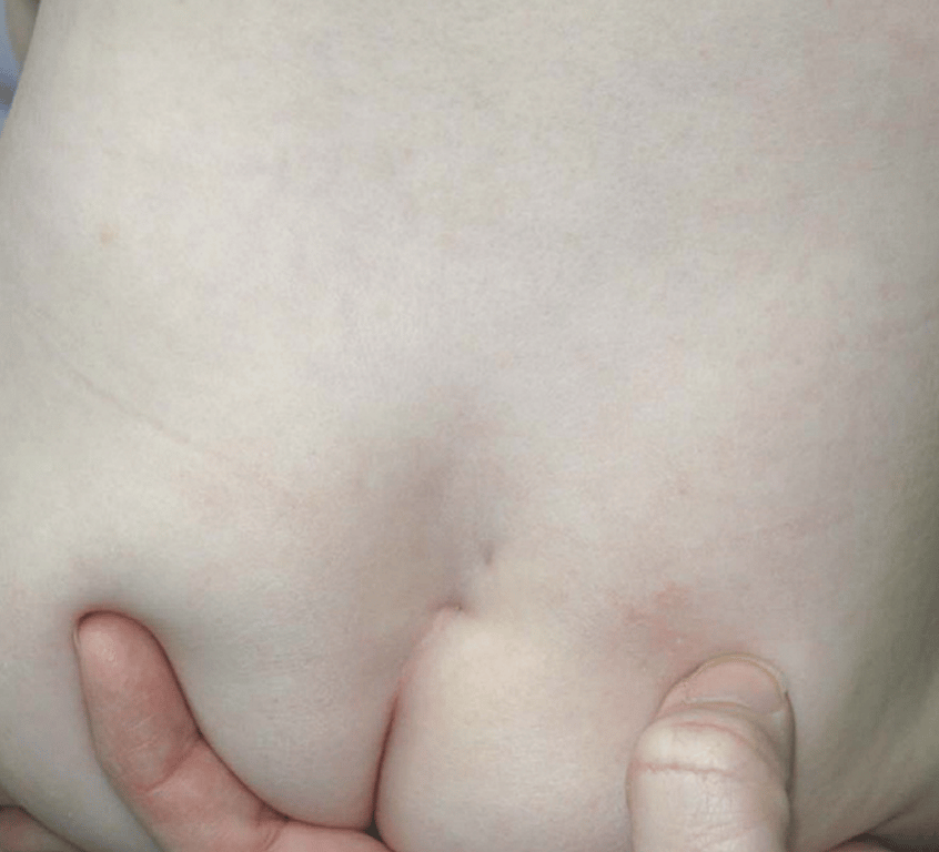
- Simple dimple
- occur in the sacral area as small depressions or pits in the skin, most with a visible floor.
- Present at birth, but sometimes not noticed until the infant’s 6 week check.
- Very common
- innocuous.
- unless they are large,(>5mm)
- located farther away from the anus,(>25mm)
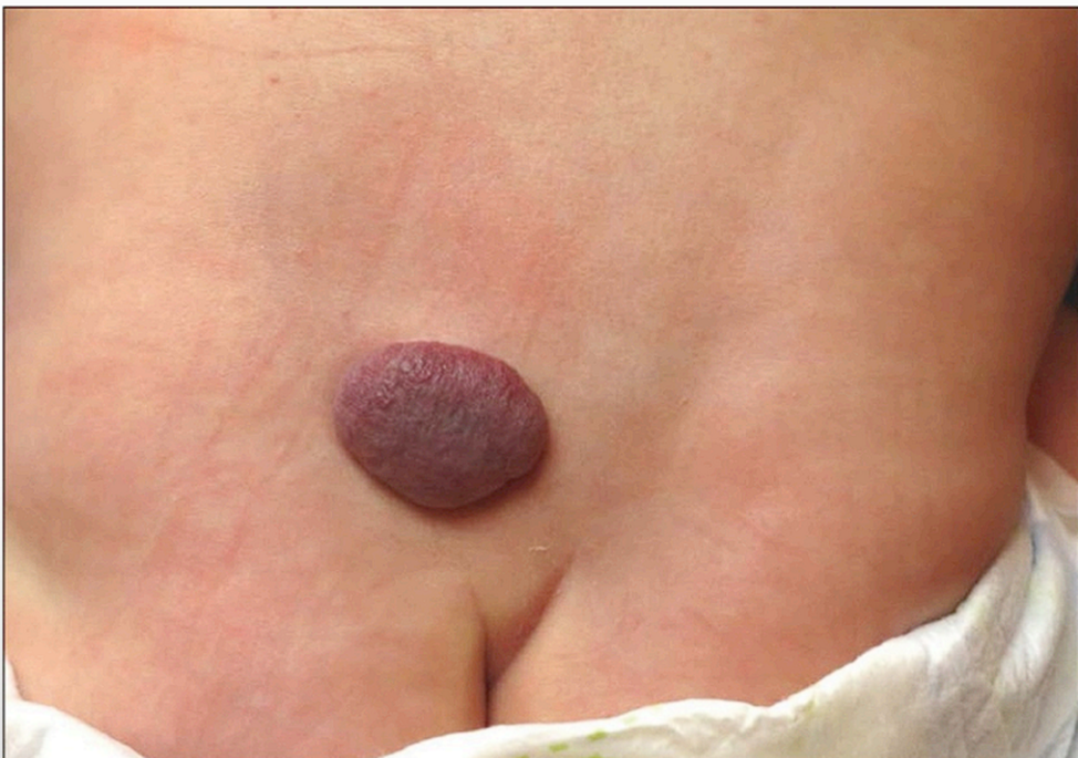
- in association with other cutaneous stigmata
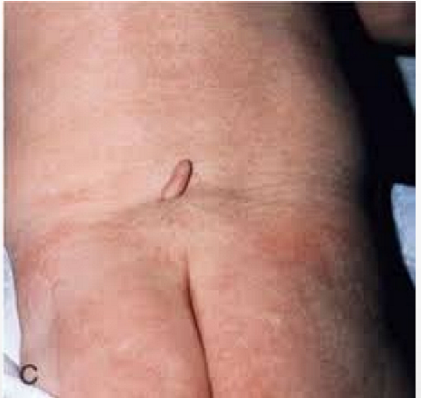
- Coccygeal pit
- A very low lying dimple with the pit pointing towards the coccygeal tip.
- Simple intergluteal dorsal dermal sinuses (dimples/pits) without other cutaneous findings do not require radiographic or surgical evaluation and treatment
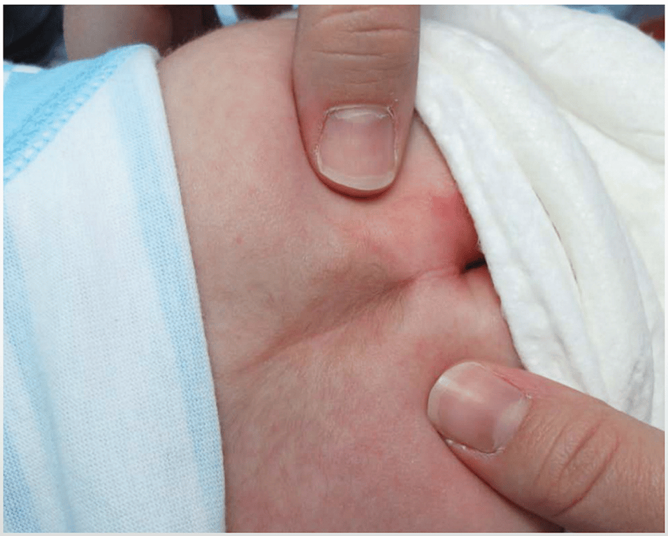
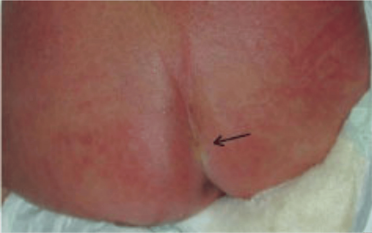
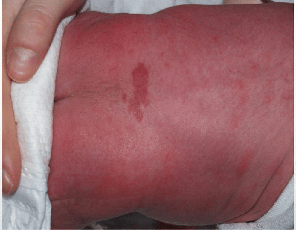
- Dimples above the gluteal cleft
- These lesions are more likely to be associated with discolouration of skin and a tuft of hair within it.
- Although the vast majority is a simple dimple, these may need to undergo further evaluation.
Causes
- Spinal bifida
- Births 1/2500
- Skin abnormalities accompany 50-80% of OSD.
- Dermal sinus tract
- Births 1/2500
- Congenital lumbosacral lipomas
- Births 1/4000
Clinical features
- Most are blind ending, just above or within the crease of the buttocks, and do not require investigation or treatment.
- Kriss et al 1998
- incidence of cutaneous stigmata in the healthy neonate study population was 4.8%.
- 207 neonates with 216 cutaneous stigmata 180 dimples(74%) None of the neonates with only a simple midline dimple had spinal dysraphism.
- 36 other cutaneous stigmata (e.g., hemangiomas, hairy patches, masses, tails) : 14 (40%) had spinal dysraphism.
- Eight (40%) of 20 atypical (>5mm, >25mm from anus, + other features) dimples were positive for spinal dysraphism
- 6 of 9 (66%) with multiple stigmata had SD.
ㅤ | Simple Dimple | Not so Simple |
Location | Within natal cleft | Above natal cleft |
ㅤ | Within 2.5 cms of anal orifice | ㅤ |
Appearance | Superficial | Deep |
ㅤ | Floor of the dimple can be seen | No floor seen (?) |
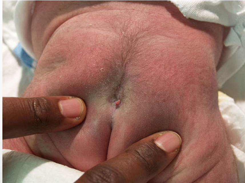
ㅤ | No discolouration | Discolouration of skin, strawberry angioma |
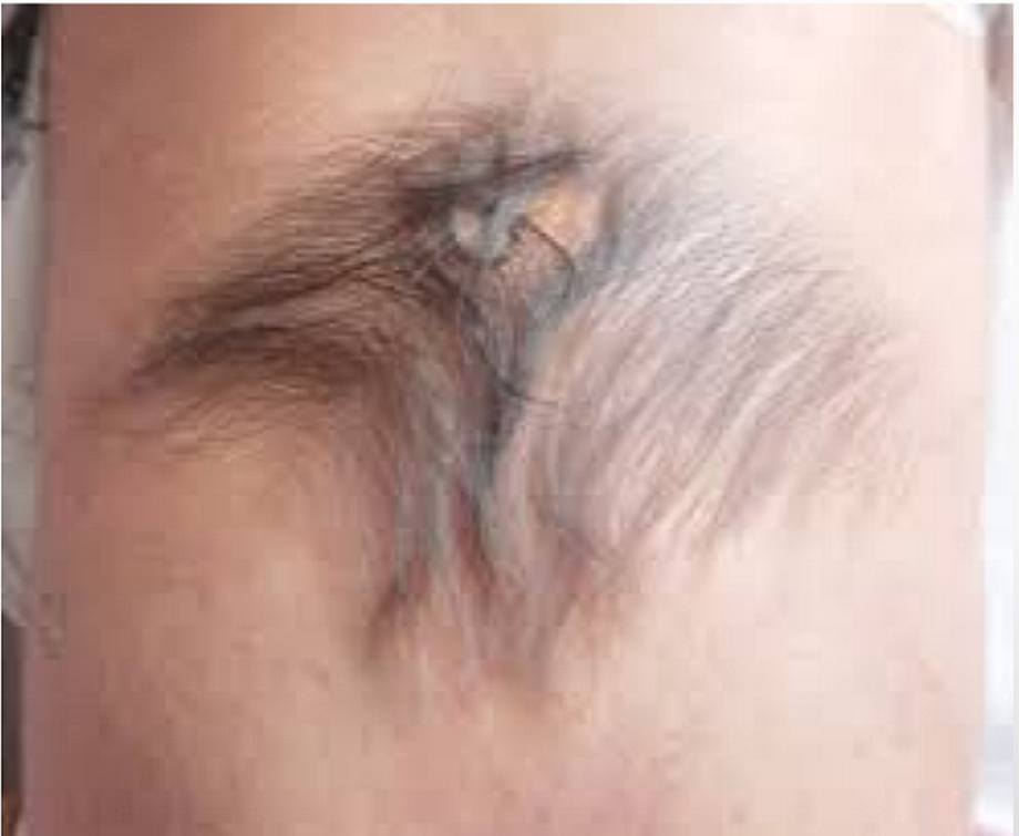
ㅤ | No tuft of hair | Tuft of hair |
ㅤ | No drainage | Fluid drainage or debris |
ㅤ | No fibrous connection or stalk Fibrous stalk directed towards coccyx tip | Fibrous stalk directed cranially |
Family History | None | Previous dimples, spina bifida, neural tube defects |
Investigation
- Refer if
- the base of the dimples cannot be visualised
- the dimple is >5mm in size
- the dimple is >2.5cm above the anal margin
- there are associated cutaneous marking, hairy patch, skin tag, or fatty lump
- True hypertrichosis, or hairs within the dimple Skin tags. Telangiectasia or hemangioma Subcutaneous mass or lump. Abnormal pigmentation. Bifurcation (fork) or asymmetry of the superior gluteal crease
- there is a duplicated gluteal cleft
- there is more than one dimple
- the dimple lies outside the sacrococcygeal region
- there are any neurological abnormalities noted
- USS
- is the initial investigation of choice, but after around 8 weeks of age (after ossification of the vertebral arches)
- MRI
- Indication
- > 5mm in depth
- > 25mm from anus
- Covered by hair
- Base not visualised
- With other cutaneous stigmata Or
- with abnormal neurology.
Reference
Images
 Made with Bullet