General
- Most common genetic cause of intellectual disability overall
- Affects 1 in every 700 infants born in the United States
Life expectancy
- 47 yrs
Pathophysiology
- Presence trisomy of chromosome 21 accounts for excessive production of specifc proteins, resulting in comorbidities such as
- Acute lymphocytic leukaemia (ALL)
- Alzheimer’s disease (AD)
Clinical features
System | Features |
CNS | Mental retardation, early dementia, hearing impairment |
Cardiac | Endocardial cushion defect: ASD, VSD, PDA, tetralogy of Fallot |
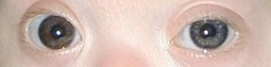
Facial | Round face, occipital/nasal flattening, Brushfield spots (speckled iris), open mouth and protruding tongue, upslanting palpebral fissure, epicanthal folds |
Hands and feet | Single transverse palmar (Simian) crease, short fingers, curved little finger (clinodactyly), sandal gap between big toe and adjacent toe |
Spine/MSK | Hypotonia, atlanto-axial instability |
GI | Duodenal atresia/stenosis, annular pancreas, omphalocele, Hirschsprung's disease, imperforate anus, tracheoesophageal fistula |
Immune | Impaired cellular immunity (more infections), autoimmune disorders, increased childhood risk of AML-M7 and ALL |
Endocrine | Hypo/hyperthyroidism, type 1 diabetes mellitus, infertility in males |
Neurological sequalae
Hypotonia
- Defined as low resting muscle tone
- 80% of newborns with DS
- Chief cause of
- Gross motor delay,
- Failure-to-thrive
- Feeding difficulties
- Tend to improve by age 3 months and resolve by 8 months of age [
- Various musculoskeletal abnormalities
Intellectual disability
- Most common genetic cause of intellectual disability,
- Can be seen in nearly all Down
- Nonverbal skills appear to develop normally, whereas language skills become deficient and never fully recover
Hearing impairment
- 47.4 and 85% of PwDS are reported to have some form of hearing impairment,
- Due to
- Middle ear effusion or other types of conductive hearing loss as the most common aetiology
- Sensorineural hearing loss accounting for 2.7–4.5%
- Prevalence increases with age and higher frequencies are the most affected
Visual impairment
- 43.3% of moderate-to-severe impairment
- Refractive errors, strabismus, epiphora, and nystagmus are the most common ocular findings
Atlantoaxial instability
- Excessive movement/ligamentous laxity between the C1 and C2 vertebrae.
- Prevalence 9.5 and 14.6%, with 1.5–3.8% of these cases becoming symptomatic, typically in the pre-teen and teen years
- Present as compressive myelopathy, which can present as a decrease in muscle strength and tone, hyperrefexia, and gait instability
- Bowel and bladder symptoms are only present in severe cases
- Don’t do radiological screening now as not all patients have it
- Instead get full hx and examine before radiological investigation
Autism spectrum disorder (ASD)
- A disorder of social communication and, in many situations, intellectual disability
Epilepsy
- Higher prevalence of epilepsy than the general population,
- Rates ranging from 1 to 13% according
- Bimodal distribution:
- The onset is prior to age one in 40%
- Infantile spasms
- Over age 30 in another 40%
- Myoclonic seizures
Moyamoya syndrome
- Prevalence of MMS is 3.8% in PwDS and as high as 9.5% in PwDS under 15 years of age
- Patients present with a bilateral infarction at onset, with 87% presenting with strokes and with a minority classified as transient ischemic attacks (TIAs). The majority of strokes were ischaemic (77% in one study) with hemorrhagic strokes being rare
Cerebral amyloid angiopathy
- Accumulation of amyloid proteins in the cerebral vessels
- Signs of microbleeds possibly associated with CAA have been reported in PwDS as young as 30
- Additional chromosome 21 which contains the gene responsible for the production of amyloid precursor protein (APP)
- Spectrum of bleeds: intraparenchymal and subarachnoid hemorrhage to cortical microbleeds and grey matter infammation
- a Convexal subarachnoid atraumatic hemorrhage in a FLAIR sequence.
- b Microinfarcts in diffusion weighted imaging.
- c Cortical superficial siderosis in T2*gradient echo sequence.
- d Lobar intracerebral hemorrhage in a CT.
- e Inflammatory reaction in FLAIR sequence.
- f Dilated perivascular spaces in T2-weighted sequence.
- g White matter hyperintensities in FLAIR sequence.
- h Cortico-subcortical microbleeds in susceptibility weighted imaging.
 Made with Bullet