- The apical ossification centre can be mistaken for a fracture;
- The synchondrosis at the base of the odontoid can be mistaken for a fracture;
- Normal synchondroses may be mistaken for fractures, especially the dentocentral synchondrosis of the axis which may be mistaken for an odontoid fracture.
- Conversely, synchondroses are biomechanically weak links and actual fractures may occur through them
- C2 is the most common vertebra injured in children.
- Vertebral bodies appear rounded-off or wedged, simulating a wedge compression fracture;
- Secondary centers of ossification at the tips of the spinous processes can be mistaken for a fracture;
- The odontoid may angulate posteriorly in 4% of children;
- C2-C3 pseudosubluxation (can be assessed with Swischuk’s line):
- Normal anterior translation that can occur between C2 and C3 and less frequently between C3 and C4 in patients younger than 8 years. May be seen in 40% of children at C2-C3 level and in 14% of children at the C3-C4 level occurs because of increased ligamentous laxity, more horizontal nature of facet joint (30° versus 60-70° in adult);
- Either anterior displacement of C2 (axis) on C3 and/or significant angulation at this level.
- May be seen in children (up to age 10 yrs) on lateral C-spine X-ray after trauma.
- Normally
- Up to age 10 yrs:
- Flexion and extension are cantered at C2–3;
- After 10 yrs:
- This moves down to C4–5 or C5–6 after age 10.
- C2 normally moves forward on C3 up to 2–3mm in paeds.
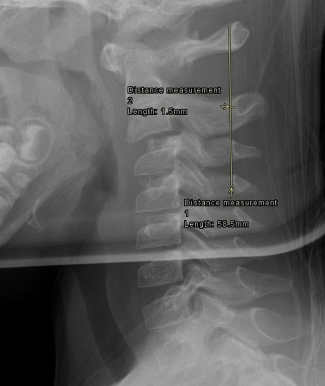
- Swischuk's line Measurement
- the line is drawn from anterior aspect of posterior arch of C1 to anterior aspect of posterior arch of C3
- the anterior aspect of posterior arch of C2 should be within 1-2 mm of this line:
- if deviated < 2 mm: it is consistent with pseudosubluxation, but this alone is insufficient to rule out a hangman fracture
- if deviated > 2 mm: it is indicative of true subluxation
- When the head is flexed, displacement is expected; may be exacerbated by spasm.
- Does not represent pathological instability.
- Fractures and dislocations are unusual in children, and when they do occur, they resemble those in adults.
- 10 cases reported between ages 4–6 yrs:
- pain was not uncommon.
- In each case, either the head or neck was flexed (sometimes minimally);
- The pseudosubluxation corrected when X-ray was repeated with head in true neutral position.
- Recommendation: treat patient for soft-tissue injury and not for subluxation.
- The ossification centre of the anterior arch of C1 may be absent in the first year of life;
- The atlanto-dens interval may be as wide as 4.5 mm and still be normal;
- The width of the prevertebral soft tissues varies widely, especially with crying, and may be mistaken for swelling;
- Horizontal facets in young children can be mistaken for a fracture
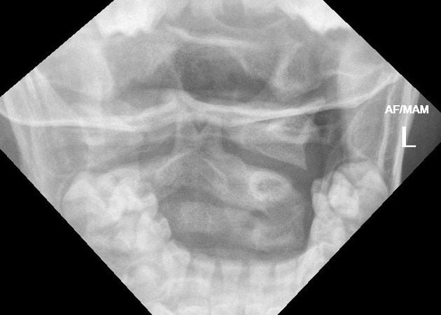
- Pseudospread of the atlas
- Aka: pseudo-Jefferson fracture
- >2mm total overlap of the two C1 lateral masses on C2 on AP open-mouth view.
- A normal overhanging of the lateral edges of the lateral masses of C1 over the lateral edges of the body of C2 seen in children.
- Due to differential speed of growth of the atlas and axis.
- This could be misdiagnosed as a Jefferson fracture, which rarely occurs prior to the teen-ages due to
- Lower weight of children,
- More flexible necks,
- Increased plasticity of skull
- Shock absorbing synchondroses of C1
- Numbers
- It is present in most children 3 mos to 4 yrs of age.
- Prevalence is 91–100% during the second year of life.
- Youngest example at 3 mos, oldest at 5.75 yrs.
- Normal total offset is
- 2mm during the first year,
- 4mm during the second,
- 6mm during the third, and decreasing thereafter.
- The maximum is 8mm.
- Trauma is not a contributing factor.
- Neck rotation can also sometimes simulate the appearance of a Jefferson fracture.
- When suspicion of a Jefferson fracture is high: thin cut CT scan parallel to and through C1 can resolve the issue of whether or not there is a fracture.
 Made with Bullet