General
- A process that starts 30 days post-conception → until 2 years of postnatal age
- exponential proliferation of the granule cell progenitors (GCP) starts at 24th gestational week and continues during postnatal age, achieving a peak at the 32nd gestational week
- This process provokes an increase in the cerebellar mass that exceeds the volume of the posterior fossa, determining a series of folding along the anterior/posterior axis and allowing the expansion of the cerebellar surface
- This foliation starts with the organization of the “anchoring centers” and the appearance of scissures that form the folia and separate the lobes.
- Cerebellum originates from the dorsal portion of the hindbrain
4 steps of cerebellum development:
- Organization of the cerebellar territory
- In the upper part of the hindbrain, the rhombomere 1
- The expression of the basic-helix-loop-helix proteins ATOH1 marks the upper rhombic lip (RL)
- The expression of the basic-helix-loop-helix proteins PTF1a marks the ventricular zone (VZ)
- In the lower portion of the hindbrain (lower RL)
- Forms
- Roof plate
- A transient pseudostratified epithelium constituted by a population of cells expressing the protein WNT1
- This layer covers the fourth ventricle roof
- Choroid plexus cells
- Production of the cerebrospinal fluid (CSF)
- Establishment of cerebellar progenitors (GABAergic and glutamatergic ones)
- Cerebellar nuclei cells are the first to be born
- Ventricular zone: Alar plate
- The ventricular zone is the neuroepithelium of the alar plate which later develops into the roof of the fourth ventricle.
- Becoming interneurons
- Derives
- Neurons
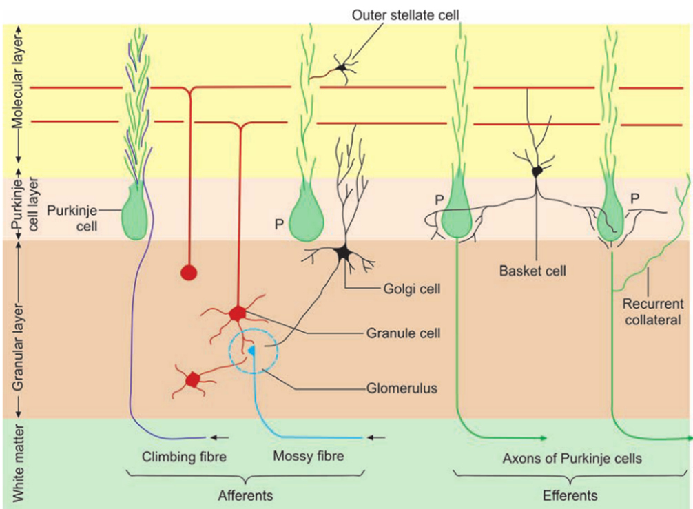
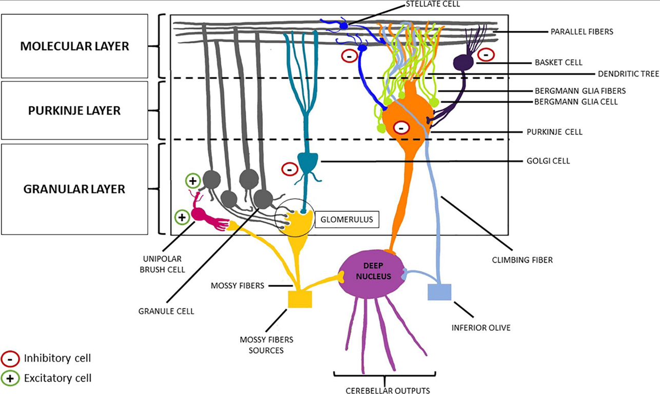
- Purkinje cells
- Two groups of PCs leave the VZ to form the so-called “Purkinje cell plate”
- Early PCs
- born in the posterior VZ
- migrate tangentially, then change orientation toward the EGL under the influence of the protein reelin secreted by the GCPs
- Late PCs
- born in the anterior VZ
- move following a radial pattern, guided by Bergmann glial fibres signalling
- Subsequently, the PCs plate reorganizes itself to form a monolayer of PCs, beneath which the IGL will locate
- The granule cells produce trophic factors necessary to develop the PCs dendritic trees.
- At 20th gestational week, the human cerebellum presents a transient cellular region called “lamina dissecans,” between the PCs layer and the IGL.
- Its function in the cerebellar development is yet unknown and it disappears by the 32nd gestational week
- Interneurons
- All inhibitory interneurons come from a common progenitor expressing a protein called PAX2 and,
- During the 3rd trimester of pregnancy
- differentiate into
- Golgi cells, that will establish in the granule layer
- Stellate and basket cells, that will take place in the molecular layer
- Glial cells
- Originate from the VZ
- Eg
- Cerebellar astrocytes
- Bergmann glia (BG)
- A small number of oligodendrocytes expressing Olig2 domain
- Glial cells are involved in numerous processes of the cerebellar development:
- cellular migration (especially the PCs)
- synapse organization
- production of neurotrophic factors
- formation of the blood-brain barrier
- Neurotransmitter: inhibitory GABAergic
- Rhomboid lip: Neural folds
- Derives
- Cerebellar nuclei neurons
- Interneurons
- Unipolar brush cells
- Especially represented in the flocculonodular lobe
- Granule layer cells
- are the glutamatergic neurons
- spring up from granule cell progenitors (GCP)
- migrates tangentially to form the external granule layer (EGL)
- following FGF8 and SHH signalling,
- goes through clonal expansion during the late pregnancy period,
- determining the formation of a six-eight cells layer
- This process produces an amount of granule neurons so large that it overcomes the cerebral cortex ones
- Later, GCPs differentiate and move inward into the cerebellar anlage to form the internal granule layer (IGL).
- During the postnatal age, the RL continues to produce granule cells and the EGL progressively disappears during the second year
- Development of the cerebellar nuclei starts with a “nuclear transitory zone” in a marginal position.
- The glutamatergic neurons follow a tangential pattern of migration and establish the GABAergic interneurons further maturation.
- The lateral nuclei develop early and project to thalamus and midbrain,
- The medial group appears later and make connections to the hindbrain
- Derivatives of the hindbrain that will form extra-cerebellar structures of the CNS also arise from the RL, like the pontine nuclei. A strict interconnection occurs between brainstem nuclei and cerebellum as the brainstem delivers proprioceptive/vestibular/auditory sensations and cortical information to the cerebellum
- Migration of the granule cells
- Formation of the cerebellar nuclei and circuitry
GABAergic ones descending from the VZ
Glutamatergic ones originating from the RL