
Early Development of the Spinal Cord
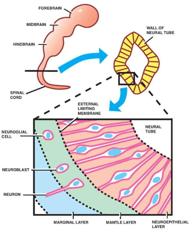
- Three layers of cells are formed from the proliferation and differentiation of the thick, pseudostratified neuroepithelium that makes up the wall of the neural tube.
- Innermost
- Neuroepithelium (or ependymal)
- A layer of ciliated columnar cells
- Lines the future ventricles and central canal.
- Gives rise to
- Neuroblasts
- Primitive neurons
- Migrate peripherally to the mantle layer
- Astrocytes and oligodendrocytes
- Migrate peripherally to the mantle and marginal layers.
- Middle
- Will later form the gray matter of the spinal cord.
- Neuroblasts in the mantle layer develop into mature neurons with cytoplasmic processes,
- Cytoplasmic processes extend peripherally to form the outermost marginal layer that later becomes the white matter of the spinal cord.
- Outer most
Neuroepithelial layer
Mantle layer
Marginal layer
- Spinal Gray Matter
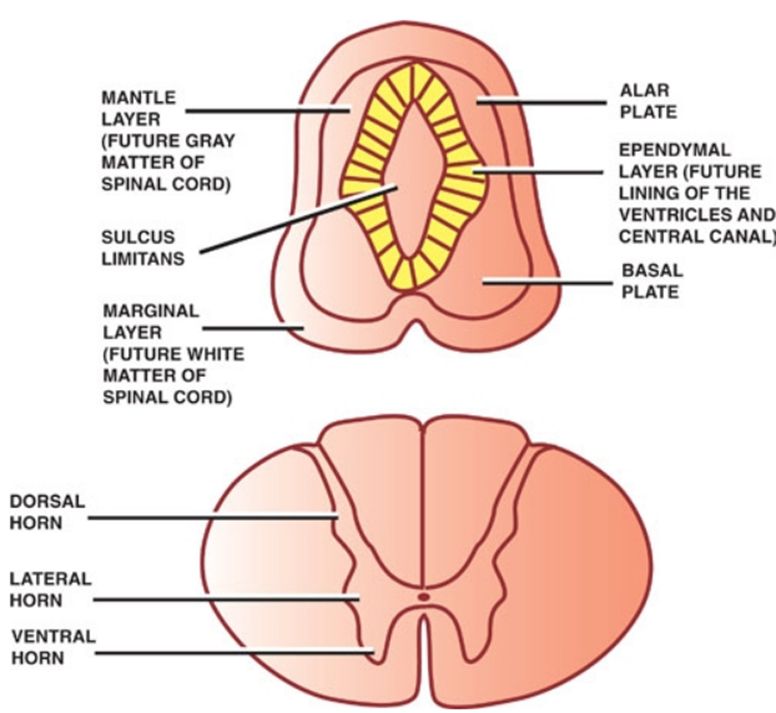
- Develops along the sides of the central cavity.
- A longitudianl groove between the dorsal and ventral aspects of the neural tube produces
- Aka: dorsal horns
- Dorsal thickening of the neural tube
- Forms the dorsal Gray columns: Sensory afferent neurons.
- Ventral and lateral thickening of the neural tube
- Contains somatic and autonomic motor neurons
- Ventral gray columns (ventral horn)
- Somatic motor neurons
- Lateral gray columns (lateral horn)
- Autonomic motor neurons
Sulcus limitans
Alar plate
Basal plate
Ventral and Dorsal Roots
- For all
- All start cranially to caudally
- Eventually the spinal nerve carries
- Somatic motor neurons
- Somatic sensory neurons
- Visceral efferent (sympathetic)
- Motor neurons
- Forms first D30
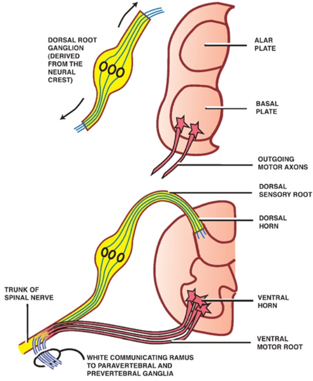
- Motor neurons in the ventral gray columns
- derived from the basal plate.
- They project axons peripherally into the ventral motor roots .
- Somatic motor neurons
- in the ventral motor roots
- join peripheral branches of the dorsal root ganglia in the region of the intervertebral foramina to form the spinal nerves.
- Sympathetic motor neurons
- in the ventral motor roots
- join the spinal nerves but exit soon after in the white communicating ramus to reach the paravertebral and prevertebral ganglia.
- Sensory neurons
- Forms second
- Cell bodies located in the dorsal root ganglia
- Derived from the neural crest.
- Pseudounipolar neurons
- Project both central and peripheral branches (axons).
- Central branches of the dorsal root ganglia
- Enter the spinal cord through the dorsal sensory roots
- Synapse in the dorsal gray column (spinothalamic tract)
- Ascend in the dorsal white column to terminate in the dorsal column nuclei (dorsal column-medial lemniscus tract).
- Neurons in the dorsal gray column and the dorsal column nuclei are derived from the alar plate.
- Peripheral branches of the dorsal root ganglia
- Enter the spinal nerves
- Course peripherally
- Terminate as sensory endings in somatic or visceral structures.
- Autonomic: sympathetic
- Forms last
- Derived from the neural crest.
Ascent of the Conus Medullaris
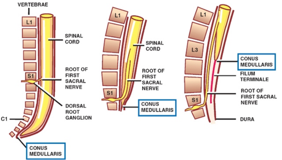
- Early stages of development, the rate of growth of the spinal cord keeps pace with that of the vertebral column; thus, the spinal nerves pass through the intervertebral foramina at their respective level of origin in the spinal cord.
- 3rd month of embryonic development:
- Rate of growth of the vertebral column > spinal cord
- End of the spinal cord assumes an increasingly higher position in relation to the vertebral column.
- Adults
- Conus medullaris is at L1
- Cervical spine:
- Each vertebral level corresponds to the level of the succeeding cord segment (i.e., the sixth cervical spine corresponds to the level of the seventh spinal cord segment)
- Upper thoracic spine:
- Difference is two segments
- Lower thoracic and upper lumber spine:
- Difference is three segments
- Filum terminale
- a long thread of pia mater that attaches the conus medullaris to the periosteum of the coccygeal vertebrae
- At what spinal level does the spinal cord end, during pre- and postnatal life?
- Prenatal life:
- 12 weeks at C5
- 15 weeks at S3
- 24 weeks at S1
- Postnatal life:
- Newborn (40 weeks) at L3
- Adult at L1–L2, end of dural and arachnoid sac at S2
S1 | 24 weeks |
L3 | 40 weeks |
L1 | Adult |