LA:
- GA
- Prone 3pins and on montreol
- Xray check level
- Midline incision bilateral strip
- Sonopet saw for Laminoplasty
- perform it roughly 1cm -1.5 cm from spinous process and angle it so that it is within the spinal canal.
- Use a sonopet saw that is blunt at the tip
- use a Swedish to feel for the canal
- Drill holes and place Codman screws (self tapping only) and lorenze plates (bent).
- Microscope in
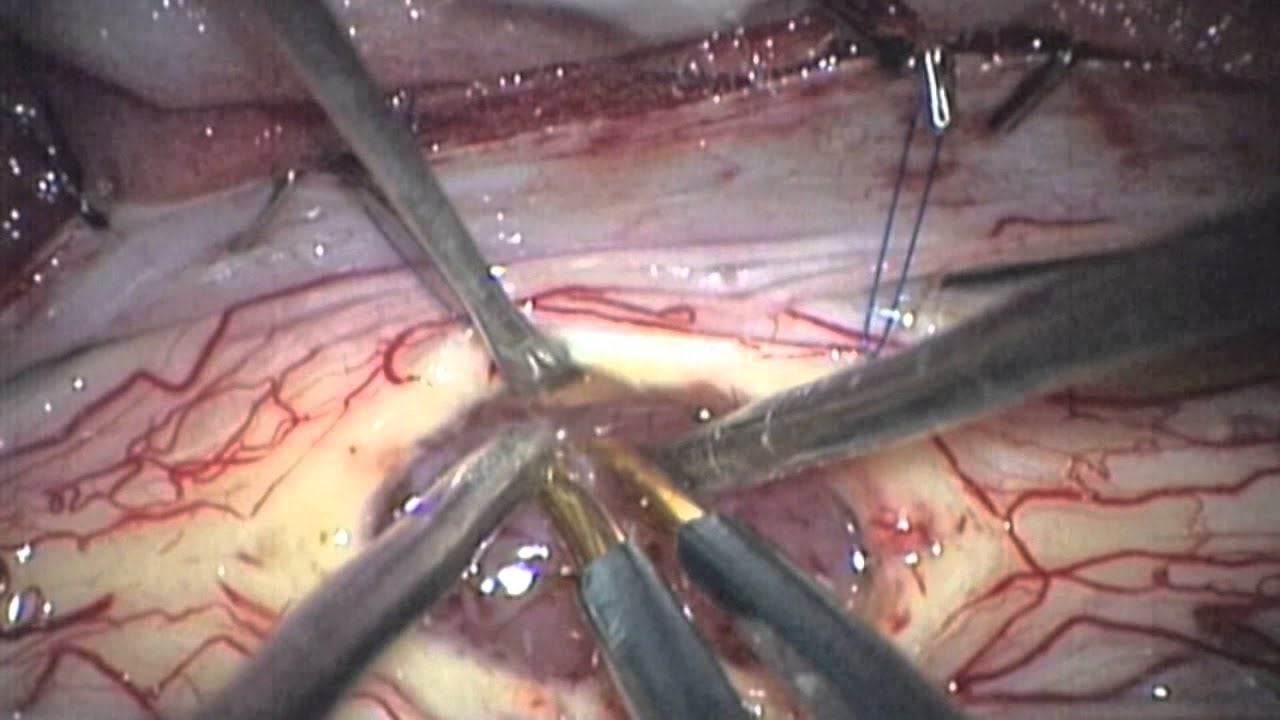
- Durotomy
- Small cut at midline with knife keeping the arachnoid intact
- Use hook or two bayonet to split the dura
- make a thin cut/line with knife to allow the dura to split in the direction of interest
- Open the arachnoid and clip it to dura
- use an orange needle to cut the arachnoid from the pia
- Find midline
- Measure from nerve root
- Neurophysiology phase reversal
- Check under high magnification and see vessels for midline raphe
- Place D wave electrode and test
- try placing it cranial if there are issues
- Try not to place the D wave wiring next to bipolar wiring
- Use knife to open midline pia
- Cut the vessel on the pial surface
- if large bipolar before cutting
- if not large vessel cut and just wash the bleeding will stop.
- Use a fenestrated tip sucker
- use micro dissector to split the whole length of the lesion so that to reduce traction on the cord when working in and around the tumour
- Take biopsy sample
- find plane around tumour with wash and micro dissector
 Made with Bullet