General
- Aka:
- ATP (Anterior to psoas approach)
- Pros
- Avoid the morbidity of the transpsoas approach by translating the incision anteriorly and dissecting around the psoas
- Lumbar plexus injury
- Minimal invasive
- Can more easily access L1/2, L2/3 than ALIF
- Avoid ALIF comorbidities
- ALIF has more
- Venous injury
- Peritoneal mobilisation → Ileus
- Avoid TLIF comorbidities
- TLIF has
- smaller cage → less endplate contact
- Risk of nerve root injury and CSF leak
- More difficult with multiple levels
- Able to perform release of ALL
- No need to reposition patient and can perform posterior fixation in same position
- Cons
- Limited exposure:
- This minimally invasive technique is suitable for exposure of L2-3, 3-4, and 4-5 levels.
- Exposure of the L4-L5 level requires ligation of the iliolumbar veins, which frequently traverse this disc space.
- Exposure of L1-2 is difficult due to 12th rib obstruction
- Increased risk to contralateral iliac vein because of oblique angle
Indication
- Foraminal stenosis
- Coronal correction for scoliosis
- Increase likelihood for arthrodesis
Relative contraindications
- Prior left sided intraperitoneal and retroperitoneal surgery
- Local radiation
Position
- Rt lateral decubitus position.
- Bed to be broken at the level of the ilium so to make the left flank working angle better.
- Slight bend of the hips to decrease the tension on the Psoas
Incision
- Xray to find level
- For L4/5
- Incision made 5cm anterior to the anterior wall of the vertebral body
- Incision made parallel to the fibers of the external abdominal oblique.
- The more anterior incision also makes the approach to the L4-5 disc space easier compared to the XLIF.
- Split the muscle in the direction of the muscle (using two fingers)
- External oblique
- Internal oblique
- Transverse abdominius
- Blunt dissect the retroperitoneal fat
- Identify anterior border of psoas
- Genitofemoral nerve overlying it
- Dock the retractor after identifying the disc space
- Working channel between Psoas and Great vessels
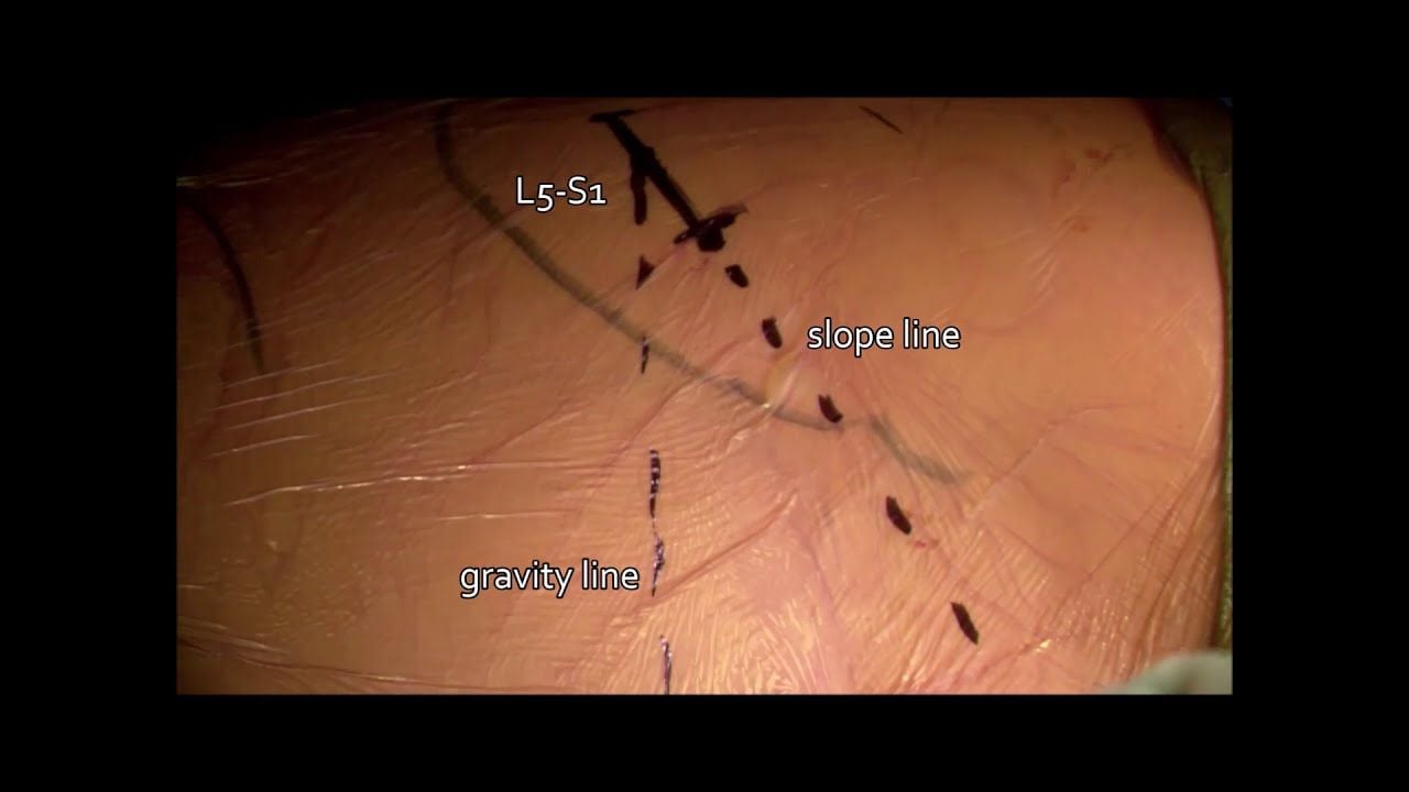
- For L5/S1
- incision is planned based on L5/S1 disc space angle
- Draw with xrays the angle of the L5/S1 disc space
- Draw a horizontal line from the L5S1 disc space
- Skin incision is 3-4 finger breaths in the line of the external oblique between the two drawn lines
- Identify retroperitoneal space
- Mobilize peritoneum and ureter medially and identify iliac vessels
- be mindful to perfrom ipsilateral discectomy to make sure implant is central
Dissection
- The external oblique, internal oblique, and transversus abdominus muscles are bluntly dissected.
- the retroperitoneal space is accessed
- the psoas muscle is identified and retracted posteriorly,
- The ureter and sympathetic plexus are retracted anteriorly.
- At this point the intervertebral space should be visible and 4 Steinman pins are used to secure the visual field surrounding the operative level of interest.
Fusion
- Once the exposure is complete, disc preparation and cage insertion are the same as for an ALIF or XLIF.
Complications
- Injury to
- Vascular structure
- Sympathetic chain
- Hypogastric plexus
- Retrograde ejaculation
- Abdominal contents
- Implant subsidence
- Abe 2017:
- Overall, 75 complications in 155 patients (48.3%);
- 69 intraoperative (44.5%)
- 7 early postoperative (4.7%)
- Only 3 patients (1.9%) had permanent damage: (All related to technical errors during disc preparation/retractor fixation)
- 1 ureteral injury
- 2 neurological injuries (nerve root and cauda equina)
- Complication types
- Endplate fracture/cage subsidence (18.7%).
- Transient psoas weakness and thigh numbness (13.5%)
- Segmental artery injury (2.6%)
- Not major vessel injury
- Surgical site infection and reoperation each 1.9%.
- OLIF vs. ExLIF Approach Comparison:
- Transpsoas group had higher rates of transient thigh/groin sensory symptoms and transient hip flexor weakness, and increased prolonged motor deficits.
- Prepsoas group had higher rates of sympathetic plexus injury (none in transpsoas) and higher rates of major vascular injury.
- Rates of bowel and urological injury were similar.
 Made with Bullet