Calcification
- Common (a sign of slow growth)
- Meningioma
- Choroid plexus papilloma
- Pineocytoma
- Uncommon
- None
Tumours that calcify
- Meningioma
- Ependymomas
- Medulloblastoma
- Subependymoma
Biphasic tumours
- Schwannoma
- Antoni A
- Antoni B
- Ganglioglioma
- Dysplastic neurons
- Neoplastic glial
- Malignant peripheral nerve sheath tumour
- Loose cellular
- Dense cellular area
Rosette forming glioneuronal tumour (RGNT)
- Uniform neurocytes forming rosettes and/ or perivascular pseudorosettes
- Astrocytic in nature and resembling pilocytic astrocytoma.
- Pilocytic astrocytoma
- Biphasic pattern with variable proportions of
- Compacted bipolar cells with rosenthal fibres and
- Loose, textured multipolar cells with micro cyst
- Papillary glioneuronal tumour
- Biphasic histological and immunophenotypic pattern with AND
- Pseudopapillary glial lining
- Interpapillary neuronal components
- Desmoplastic infantile ganglioglioma / desmoplastic infantile astrocytoma
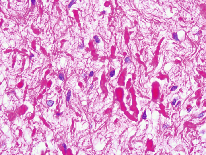
Rosenthal Fibers
- Intracytoplasmic aggregates of glial fibrillary acidic protein (GFAP) and chaperone proteins.
- They are bright eosinophilic in H&E-stained sections and cork-screw-like or beaded.
- Eosinophilic granular bodies are related to Rosenthal fibers and they often occur together.
- Found in
- Pilocytic astrocytoma
- Grade I ganglioglioma
- Grade II pleomorphic xanthoastrocytoma,
- Alexander disease
- Reactive gliosis (piloid gliosis)
- Particularly around chronic lesions in the hypothalamus, spinal cord or cerebellum (e.g., craniopharyngioma, AVM, syrinx, or granulomatous inflammation).
What are the small, round, blue cell tumors of childhood?
- Neuroblastomas
- Chondrosarcoma
- Rhabdomyosarcoma
- Lymphoma
- Ewing sarcoma
 Made with Bullet