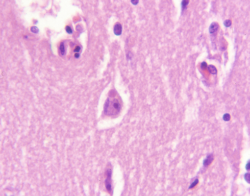
Bunina bodies
- Eosinophilic granular cytoplasmic deposits seen within anterior horn cells
- Seen in Amyotrophic lateral sclerosis
Coiled bodies
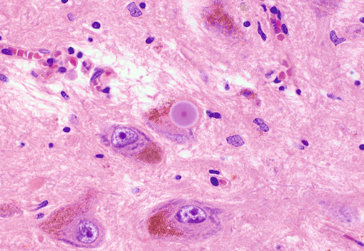
Lewy bodies
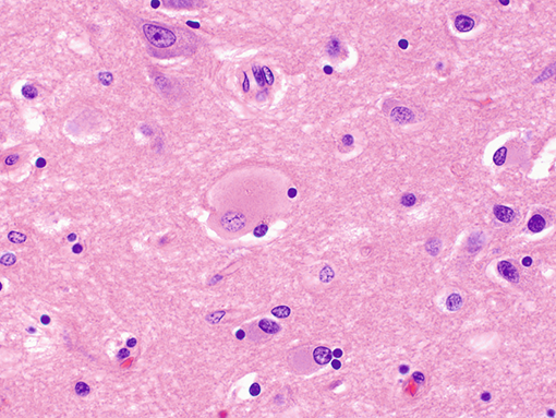
- Single to multiple intracytoplasmic inclusions composed of round, eosinophilic central material surrounded by prominent pale haloes; found within brain stem neurons and are α-synuclein immunoreactive. Other histological features of Parkinson’s disease include Lewy neurites, cortical Lewy bodies (lack prominent haloes; not the dominant feature), pale bodies and pigmented neuron loss within the substantia nigra and locus coeruleus. In Lewy body dementia, the prominent feature is cortical Lewy bodies and spongiotic change in cortical layers I-III (with senile plaques, neurofibrillary tangles, and Hirano bodies) although classical Lewy bodies may be found in substantia nigra.
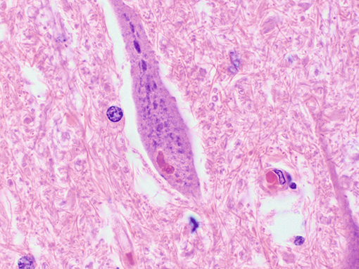
Negri bodies
- These are present in 70% of cases of rabies virus encephalitis and are ovoid/round eosinophilic cytoplasmic neuronal inclusions, predominantly found in hippocampal pyramidal neurons, brainstem/deep nuclei and Purkinje cells of cerebellum.
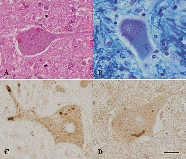
Pick disease
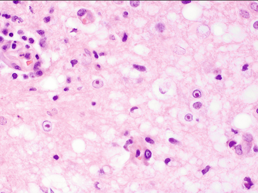
- Neurodegenerative disease with circumscribed frontotemporal cortical and subcortical degeneration characterized clinically by abnormalities in behavior, speech, and language. Intraneuronal accumulation of tau (Pick bodies; bottom) and neurons with swollen eosinophilic cytoplasm (Pick cells/“balloon” cells; center) histologically.
Hirano bodies
- Hirano bodies are hypereosinophilic rod-shaped structures typically found in the neuropil next to neurons. The may be present (but are not pathognomonic) in Alzheimer’s disease.
Cowdry type A intranuclear inclusions
- These are sparse, intranuclear inclusions associated with HSV, VZV and CMV infection (owl’s eye).
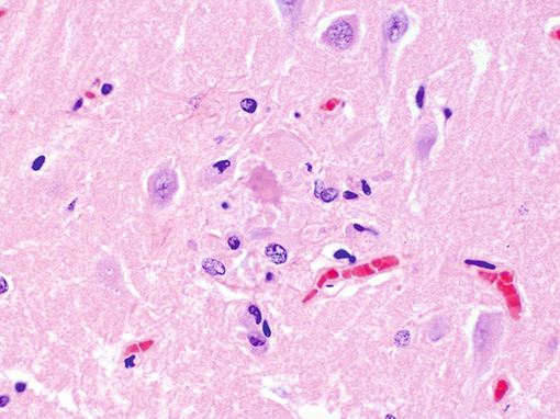
Neuritic plaque
- Neuritic (senile) plaques are extracellular deposits of Aβ-amyloid peptide (abnormal degradation of amyloid precursor protein) in the neocortex; the presence of degenerating “dystrophic” neurites (axons or dendrites) with an amyloid core characterizes a neuritic plaque, and the deposition of amyloid without dystrophic neurites is considered a diffuse plaque—both are seen in Alzheimer’s disease.
Schiller duvel Bodies
- Yolk sac tumours
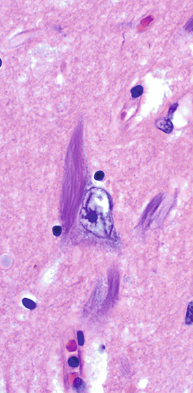
Neurofibrillary tangles
- Flame-shaped filamentous cytoplasmic structures that frequently take the shape of pyramidal neurons, and are classically seen in Alzheimer’s disease. They are neuronal cytoplasmic accumulations of ubiquitinated, hyperphosphorylated tau protein found first in limbic structures (hippocampus, amygdala, entorhinal cortex) followed by involvement of the neocortex (especially the association cortices).
 Made with Bullet